. 2003 Apr;77(8):4827-35.
doi: 10.1128/jvi.77.8.4827-4835.2003.

Complexes of poliovirus serotypes with their common cellular receptor, CD155

Affiliations

Complexes of poliovirus serotypes with their common cellular receptor, CD155

Yongning He et al. J Virol. 2003 Apr.

Abstract

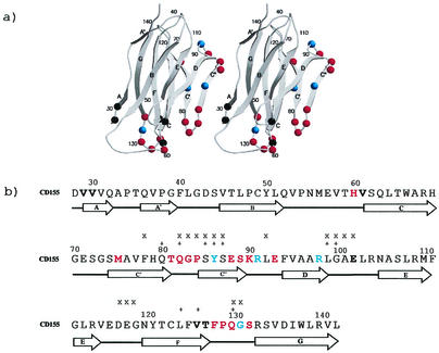
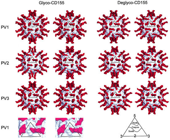
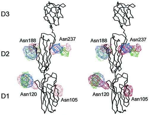
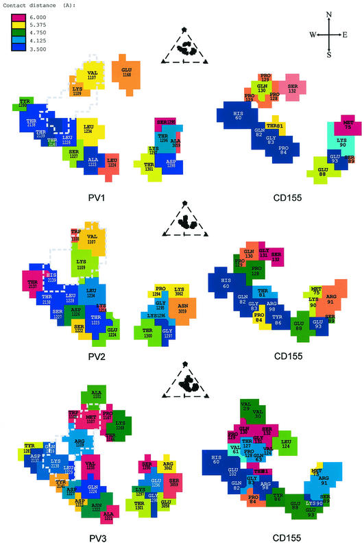
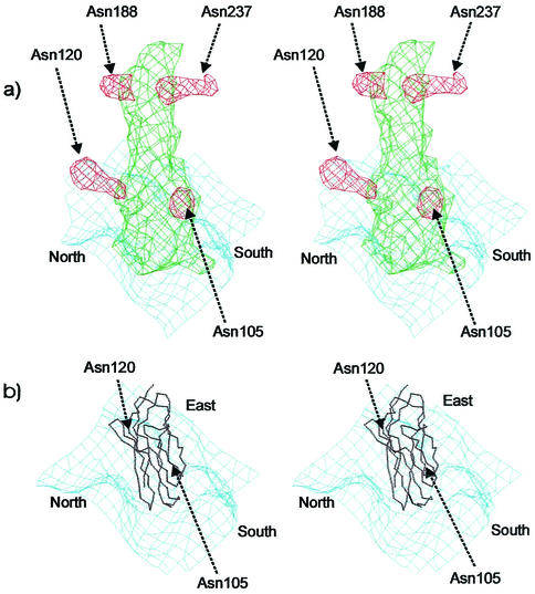
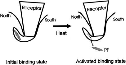
Structures of all three poliovirus (PV) serotypes (PV1, PV2, and PV3) complexed with their cellular receptor, PV receptor (PVR or CD155), were determined by cryoelectron microscopy. Both glycosylated and fully deglycosylated CD155 exhibited similar binding sites and orientations in the viral canyon for all three PV serotypes, showing that all three serotypes use a common mechanism for cell entry. Difference maps between the glycosylated and deglycosylated CD155 complexes determined the sites of the carbohydrate moieties that, in turn, helped to verify the position of the receptor relative to the viral surface. The proximity of the CD155 carbohydrate site at Asn105 to the viral surface in the receptor-virus complex suggests that it might interfere with receptor docking, an observation consistent with the properties of mutant CD155. The footprints of CD155 on PV surfaces indicate that the south rim of the canyon dominates the virus-receptor interactions and may correspond to the initial CD155 binding state of the receptor-mediated viral uncoating. In contrast, the interaction of CD155 with the north rim of the canyon, especially the region immediately outside the viral hydrophobic pocket that normally binds a cellular "pocket factor," may be critical for the release of the pocket factor, decreasing the virus stability and hence initiating uncoating. The large area of the CD155 footprint on the PV surface, in comparison with other picornavirus-receptor interactions, could be a potential limitation on the viability of PV escape mutants from antibody neutralization. Many of these are likely to have lost their ability to bind CD155, resulting in there being only three PV serotypes.


Figures

FIG. 1.
FIG. 1.
(a) Ribbon diagram of CD155 domain D1. Residues in the virus-receptor binding interfaces are shown as colored spheres. Residues identified as being in the interface in all three serotypes are shown in red, residues identified in two serotypes are shown in blue, and residues identified in only one serotype are shown in black. Secondary structure elements are identified by A, B, C, C′, C", D, E, F, and G. Residues are numbered at strategic positions. (b) CD155 residues in the virus-receptor binding interface. Residues are colored as in Fig. 1a. The residues that are important for receptor binding, as indicated by mutagenesis studies, are marked by “x” (6, 17) and “+” (9, 30, 34).
FIG. 2.
FIG. 2.
Stereoviews of cryoEM reconstructions of PV1, -2, and -3 complexed with glycosylated and fully deglycosylated CD155. Viral surfaces and CD155 are shown in gray and red, respectively. Resolutions of the different reconstructions are given in Table 1. Also shown (bottom left) is an enlarged, surface-shaded representation of the icosahedral asymmetric unit showing the complex of glycosylated CD155 with PV1. At the bottom right is an explanation of the north and south notation used to describe the canyon topology.
FIG. 3.
FIG. 3.
Stereoviews of superposition of the carbohydrate site difference densities obtained by subtracting the deglycosylated from the glycosylated CD155 densities. The carbohydrate densities are colored in red, green, and blue for those derived from the PV1, PV2, and PV3 complexes, respectively. The Cα backbone of CD155 is shown in black.
FIG. 4.
FIG. 4.
Footprints of CD155 on the surfaces of PV1 (top), PV2 (middle), and PV3 (bottom). Each figure is viewed in the same direction as those shown in Fig. 2, which is down an icosahedral twofold axis. The viral residues in the footprints are colored according to their nearest approach to a CD155 atom. The projections of the pocket factor to the CD155 footprints are outlined with a dashed line. The relative position of the CD155 footprint in the viral asymmetric unit is shown at the top of each panel. Shown at the top right is a compass to explain the notation used to describe the parts of the canyon.
FIG. 5.
FIG. 5.
(a) Stereoviews of the carbohydrate difference density (red) of domain D1 and D2 of the PV1-CD155 complex. The deglycosylated CD155 density and the viral surface are shown in green and blue, respectively. (b) The Cα backbone of CD155 D1 is shown in black. The orientation of the canyon is marked as north, south, and east.
FIG. 6.
FIG. 6.
Model of receptor-mediated PV uncoating. The south wall of the canyon is probably responsible for forming the initial binding state of the PV-CD155 complex. The interaction between the north wall of the canyon and CD155 immediately above the hydrophobic binding pocket helps the release of the pocket factor, thus destabilizing the virus. Heating increases the structural “breathing” of the virus, thus providing the conformational change required for forming the “activated state” of the PV-CD155 complex.

References

    1. Arita, M., S. Koike, J. Aoki, H. Horie, and A. Nomoto. 1998. Interaction of poliovirus with its purified receptor and conformational alteration in the virion. J. Virol. 72:3578-3586. - PMC - PubMed
    1. Baker, T. S., and R. H. Cheng. 1996. A model-based approach for determining orientations of biological macromolecules imaged by cryoelectron microscopy. J. Struct. Biol. 116:120-130. - PubMed
    1. Baker, T. S., N. H. Olson, and S. D. Fuller. 1999. Adding the third dimension to virus life cycles: three-dimensional reconstruction of icosahedral viruses from cryo-electron micrographs. Microbiol. Mol. Biol. Rev. 63:862-922. - PMC - PubMed
    1. Belnap, D. M., B. M. McDermott, Jr., D. J. Filman, N. Cheng, B. L. Trus, H. J. Zuccola, V. R. Racaniello, J. M. Hogle, and A. C. Steven. 2000. Three-dimensional structure of poliovirus receptor bound to poliovirus. Proc. Natl. Acad. Sci. USA 97:73-78. - PMC - PubMed
    1. Bernhardt, G., J. A. Bibb, J. Bradley, and E. Wimmer. 1994. Molecular characterization of the cellular receptor for poliovirus. Virology 199:105-113. - PubMed

Publication types

Associated data

LinkOut - more resources