. 2003 Apr;77(8):4858-66.
doi: 10.1128/jvi.77.8.4858-4866.2003.

Binding of adenovirus capsid to dipalmitoyl phosphatidylcholine provides a novel pathway for virus entry

Affiliations

Binding of adenovirus capsid to dipalmitoyl phosphatidylcholine provides a novel pathway for virus entry

Larissa Balakireva et al. J Virol. 2003 Apr.

Abstract

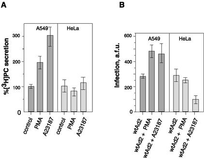
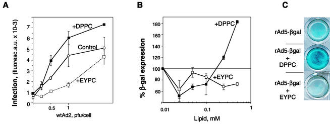
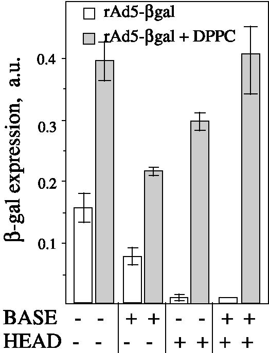
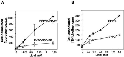
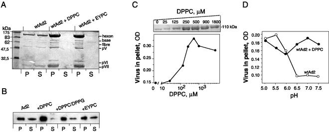
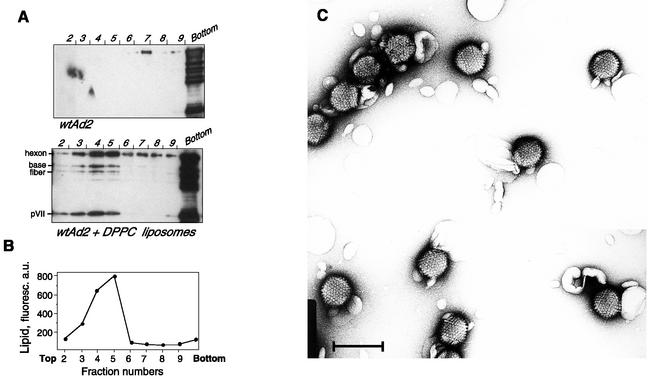
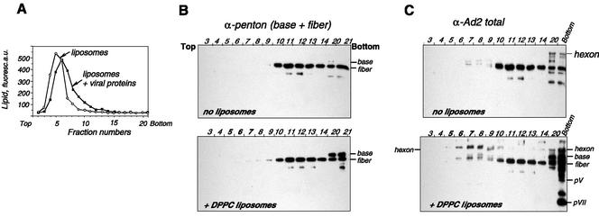
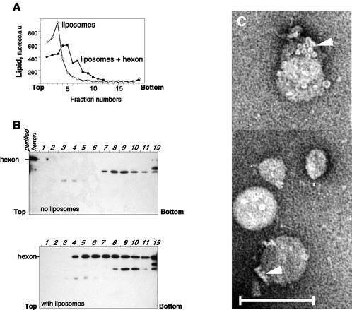
Adenovirus (Ad) is an airborne, nonenveloped virus infecting respiratory epithelium. To study the mechanism of Ad entry, we used alveolar adenocarcinoma A549 cells, which have retained the ability of alveolar epithelial type II cells to synthesize the major component of pulmonary surfactant, disaturated phosphatidylcholine. Stimulation of phosphatidylcholine secretion by calcium ionophore or phorbol ester augmented the susceptibility of these cells to Ad. Both Ad infection and recombinant-Ad-mediated transfection increased in the presence of dipalmitoyl phosphatidylcholine (DPPC) liposomes in culture medium. Importantly, in the presence of DPPC liposomes, virus penetrates the cells independently of virus-specific protein receptors. DPPC vesicles bind Ad and are efficiently incorporated by A549 lung cells, serving as a virus vehicle during Ad penetration. To identify the viral protein(s) mediating Ad binding, a flotation of liposomes preincubated with structural viral proteins was employed, showing that the only Ad protein bound to DPPC vesicles was a hexon. The hexon preserved its phospholipid-binding properties upon purification, confirming its involvement in virus binding to the phospholipid. Given that disaturated phosphatidylcholine not only covers the inner surface of alveoli in the lungs but also reenters alveolar epithelium during lung surfactant turnover, Ad binding to this phospholipid may provide a pathway for virus entry into alveolar epithelium in vivo.


Figures

FIG. 1.
FIG. 1.
Effect of A23187 and PMA on phosphatidylcholine secretion and Ad2 infectivity. (A) A549 and HeLa cells prelabeled with [3H]choline were incubated with A23187 or PMA, and 2 h later, the amount of [3H]phosphatidylcholine ([3H]PC) in culture medium was measured as described in Materials and Methods. Data are percentages of [3H]PC secreted under control conditions taken as 100% (0.02% dimethyl sulfoxide or 15 nM α-PMA) and in the presence of 5 μM A23187 or 15 nM PMA, respectively. (B) A549 and HeLa cells were exposed to A23187 (5 μM) or PMA (15 nM) for 1 h, followed by the addition of Ad2 (MOI = 0.5). One hour later, virus was removed and cells were cultured for an additional 20 h. The synthesis of late viral proteins was estimated after immunostaining as described in Materials and Methods. Data are given in arbitrary fluorescence units (a.f.u.) associated with the cell monolayer. Bars indicate the means ± standard errors (n = 6).
FIG. 2.
FIG. 2.
DPPC liposomes enhance the infectivity of wild-type Ad2 (wtAd2) and augment the efficiency of rAd-mediated transfection. (A) Serially diluted Ad2 was added to A549 cells together with EYPC or DPPC liposomes (0.62 mM). At the end of a 45-min incubation period at 37°C, the virus-liposome suspension was removed and cells were cultured for an additional 20 h before being processed for indirect immunofluorescence staining. Bars indicate the means ± standard errors (n = 2). Curves representative of three independent experiments are shown. (B and C) A549 cells were transduced with rAd5-βgal (MOI = 50) in the presence of increasing concentrations of liposomes (0.025 to 0.625 mM) for 45 min at 37°C. Virus was removed, and the expression of β-galactosidase was measured 24 h later, either as the optical density of the cell monolayer measured at 630 nm (B) or after in situ X-Gal staining of methanol-fixed cells (C). Bars in panel B indicate the means ± standard errors (n = 2).
FIG. 3.
FIG. 3.
DPPC liposomes mediate Ad entry via a receptor-independent pathway. (A) A549 cells were preincubated with the recombinant viral proteins either penton base (80 μg/ml) or fiber head domain (8 μg/ml) at 4°C for 1 h. DPPC liposomes (0.5 mg/ml) and rAd5-βgal (at an MOI of 100) were then added for 45 min. The expression of β-galactosidase was measured 24 h later. Error bars represent the standard deviations around the means for three independent measurements. a.u., arbitrary units.
FIG. 4.
FIG. 4.
Liposome uptake by A549 cells. A549 cells were incubated with DPPC-NBD-PE or EYPC-NBD-PE liposomes (A) or with [3H]choline-loaded DPPC or EYPC liposomes (B) for 1 h and thoroughly rinsed with PBS, and cell-associated fluorescence or radioactivity was measured. Bars indicate the means ± standard errors (n = 2). a.u., arbitrary units.
FIG. 5.
FIG. 5.
Cosedimentation of purified Ad with liposomes. (A) Liposomes (100 μg) were incubated with 10 μg of Ad2 in 100 μl of HBS, pH 7.0, and processed as described in Materials and Methods. (B) Similar analyses performed at lower concentrations of lipid (8 μg/100 μl) and Ad2 (2 μg/100 μl) were revealed by Western blotting with polyclonal anti-Ad2 antibodies (only the hexon band is shown). (C) Ad2 (3.7 μg) was incubated with increasing amounts of DPPC liposomes and centrifuged. The pellet fractions were analyzed by SDS-PAGE. The amount of Ad precipitated by DPPC liposomes was estimated by densitometric scanning of the hexon protein after Coomassie staining (upper insert). OD, optical density. (D) Effect of pH on the formation of Ad2-liposome complexes. Ad2 (100 μg/ml) was incubated with DPPC liposomes (0.5 mM) in 50 mM morpholineethanesulfonic acid-OH (pH below 6) or 50 mM HEPES-OH (pH above 6.0) and recovered by centrifugation, and its amount was estimated as shown in panel B. The representative data from at least two independent experiments are shown.
FIG. 6.
FIG. 6.
Coflotation of Ad with DPPC liposomes. (A) Ad2 (9 μg) was incubated with DPPC-NBD-PE liposomes (20 μg) for 15 min. (B) Next, it was processed for Western blot analysis as described in Materials and Methods. a.u., arbitrary units. (C) Electron microscopy of the Ad2-DPPC liposome mixture was done. Scale bar, 100 nm.
FIG. 7.
FIG. 7.
Identification of viral proteins bound to DPPC liposomes. Soluble viral proteins were incubated with DPPC-NBD-PE liposomes in HBS as described in Materials and Methods. Gradient fractions were analyzed for the presence of lipid by measuring NBD-PE fluorescence (in arbitrary units [a.u.]) (A). The protein content of each fraction was analyzed by Western blot analysis using anti-penton (B) or anti-Ad2 (C) antibody. Gels representative of three independent experiments are shown.
FIG. 8.
FIG. 8.
Coflotation of purified hexon with DPPC liposomes. Hexon purified by DEAE-Sepharose chromatography was mixed with DPPC liposomes and centrifuged on a sucrose density gradient (6 to 40%), and gradient fractions were analyzed for NBD-PE fluorescence (A) or by Western blotting with anti-Ad2 antibody (B). a.u., arbitrary units. (C) Electron microscopy analysis of fraction 9 (with liposomes) was carried out after negative staining. Note groups-of-nine hexons (arrowheads) on the liposome surface. Scale bar, 100 nm.

References

    1. Akalu, A., H. Liebermann, U. Bauer, H. Granzow, and W. Seidel. 1999. The subgenus-specific C-terminal region of protein IX is located on the surface of the adenovirus capsid. J. Virol. 73:6182-6187. - PMC - PubMed
    1. Arcasoy, S. M., J. Latoche, M. Gondor, S. C. Watkins, R. A. Henderson, R. Hughey, O. J. Finn, and J. M. Pilewski. 1997. MUC1 and other sialoglycoconjugates inhibit adenovirus-mediated gene transfer to epithelial cells. Am. J. Respir. Cell. Mol. Biol. 17:422-435. - PubMed
    1. Benson, S. D., J. K. Bamford, D. H. Bamford, and R. M. Burnett. 2002. The X-ray crystal structure of P3, the major coat protein of the lipid-containing bacteriophage PRD1, at 1.65 Ä resolution. Acta Crystallogr. Sect. D Biol. Crystallogr. 58:39-59. - PubMed
    1. Benson, S. D., J. K. H. Bamford, D. H. Bamford, and R. M. Burnett. 1999. Viral evolution revealed by bacteriophage PRD1 and human adenovirus coat protein structures. Cell 98:825-833. - PubMed
    1. Bergelson, J. M., J. A. Cunningham, G. Droguett, E. A. Kurt-Jones, A. Krithivas, J. S. Hong, M. S. Horwitz, R. L. Crowell, and R. W. Finberg. 1997. Isolation of a common receptor for coxsackie B viruses and adenoviruses 2 and 5. Science 275:1320-1323. - PubMed

Publication types

LinkOut - more resources