. 2003 Apr;77(8):4888-98.
doi: 10.1128/jvi.77.8.4888-4898.2003.

Membrane association of VP22, a herpes simplex virus type 1 tegument protein

Affiliations

Membrane association of VP22, a herpes simplex virus type 1 tegument protein

Michael J Brignati et al. J Virol. 2003 Apr.

Abstract

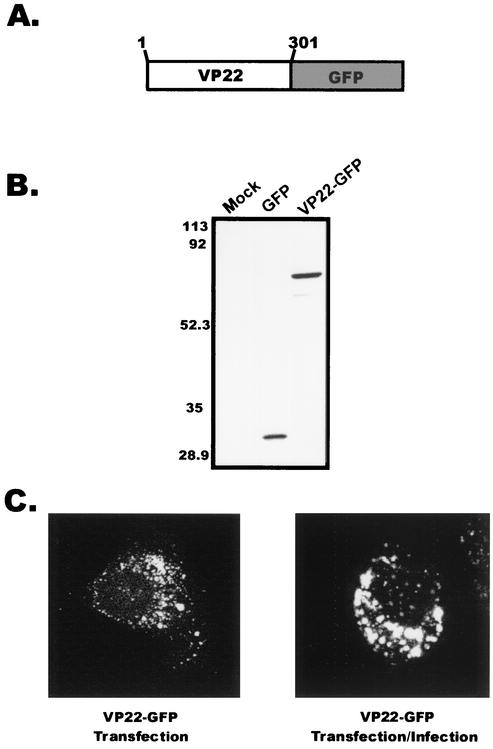
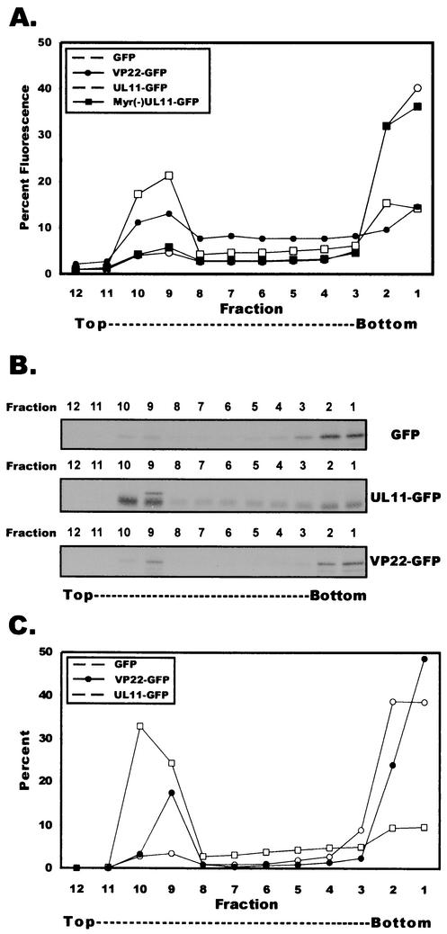
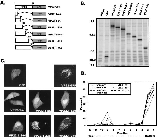
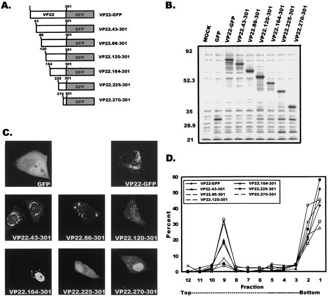
Tegument proteins of herpes simplex virus type 1 (HSV-1) are hypothesized to contain the functional information required for the budding or envelopment process proposed to occur at cytoplasmic compartments of the host cell. One of the most abundant tegument proteins of HSV-1 is the U(L)49 gene product, VP22, a 38-kDa protein of unknown function. To study its subcellular localization, a VP22-green fluorescent protein chimera was expressed in transfected human melanoma (A7) cells. In the absence of other HSV-1 proteins, VP22 localizes to acidic compartments of the cell that may include the trans-Golgi network (TGN), suggesting that this protein is membrane associated. Membrane pelleting and membrane flotation assays confirmed that VP22 partitions with the cellular membrane fraction. Through truncation mutagenesis, we determined that the membrane association of VP22 is a property attributed to amino acids 120 to 225 of this 301-amino-acid protein. The above results demonstrate that VP22 contains specific information required for targeting to membranes of acidic compartments of the cell which may be derived from the TGN, suggesting a potential role for VP22 during tegumentation and/or final envelopment.


Figures

FIG. 1.
FIG. 1.
Expression of VP22 within A7 cells. (A) Construction of VP22-GFP. VP22 from the HSV-1 KOS genome was cloned as a C-terminal fusion to the GFP protein. (B) Western blot analysis. A7 melanoma cells were transfected with the indicated plasmids, and cell lysates were separated by SDS-PAGE. Proteins transferred to nitrocellulose membranes were probed with a polyclonal antibody specific for GFP. (C) Subcellular localization of VP22. Plasmid DNA encoding VP22-GFP was transfected into A7 cells as in the experiment for which results are shown in panel B, and localization of the VP22-GFP chimera was observed in the absence or presence of HSV-1 infection. Fluorescence was visualized by live-cell confocal microscopy at approximately 32 h posttransfection (transfection only) or 32 h posttransfection-infection (24 h posttransfection plus 8 h postinfection).
FIG. 2.
FIG. 2.
Subcellular localization of VP22 in A7 cells. (A to F) Subcellular localization of VP22 relative to those of cellular markers for the TGN. Immunofluorescence was performed on A7 cells that were transfected with the plasmid encoding VP22-GFP. After 20 h posttransfection, cells were fixed with paraformaldehyde and permeabilized with Triton X-100. In panels A to C, cells were labeled with a monoclonal antibody against AP-1, which was detected by a rabbit anti-mouse antibody conjugated to TRITC. Panels A and B are the same field viewed by confocal microscopy with the appropriate wavelength to excite GFP (A) or TRITC (B), and these images were digitally combined to produce the image in panel C. In panels D to F, cells were labeled with a polyclonal sheep antibody against TGN-46, which was detected by a rabbit anti-sheep antibody conjugated to Texas Red. Panels D and E are the same field viewed by confocal microscopy with the appropriate wavelength to excite GFP (D) or Texas Red (E), and these images were digitally combined to produce the image in panel F. (G to I) Subcellular localization of VP22 relative to the acidic compartments of the cell. Live-cell fluorescence was performed on A7 cells expressing VP22-GFP and stained with LysoTracker Red. Panels G and H are the same field viewed by confocal microscopy with the appropriate wavelength to excite GFP (G) or LysoTracker (H), and these images were digitally combined to produce the image in panel I.
FIG. 3.
FIG. 3.
VP22 pellets with cellular membranes. A7 cells were transfected with the indicated plasmids, harvested at 20 h posttransfection, and incubated in hypotonic buffer. Swollen cells were disrupted by Dounce homogenization, and nuclei were removed by low-speed centrifugation. In order to separate membrane-bound entities from soluble forms, the supernatants underwent centrifugation at 100,000 × g. As a control, replica samples were incubated with 0.5% Triton X-100 prior to centrifugation to solubilize cellular membranes. Following centrifugation, soluble and pellet fractions were collected and analyzed for the presence of GFP chimeras by fluorometry. The fraction pelleted was calculated by dividing the amount pelleted by the total amount of protein in the soluble and membrane fractions (see Materials and Methods). Error bars, standard deviations for three independent experiments.
FIG. 4.
FIG. 4.
Membrane flotation of VP22. (A) Membrane flotation of VP22 assayed by fluorometry. A7 cells were transfected with the indicated plasmids, harvested, and incubated in hypotonic buffer. Swollen cells were disrupted by Dounce homogenization, and nuclei were removed by low-speed centrifugation. Membrane flotation gradient centrifugation was then performed on postnuclear supernatants as described in Materials and Methods. Gradient fractions were analyzed for the presence of GFP chimeras by fluorometry. The percentage of fluorescence for a given fraction was determined by dividing the fluorescence of a given fraction by the sum total fluorescence of all fractions (see Materials and Methods). Fluorometric results are depicted graphically. (B) Membrane flotation of VP22 assayed by immunoprecipitation. A7 cells were transfected with the indicated plasmids and were metabolically labeled with EXPRE35S35S protein labeling mix for 2.5 h. Membrane flotation gradient centrifugation was performed as described above. Gradient fractions were collected, diluted in RIPA buffer, and incubated with a polyclonal goat antibody specific for GFP.Immunoprecipitated fractions were separated by SDS-PAGE and visualized by autoradiography. (C) Quantification of membrane flotation. SDS-PAGE gels described above were analyzed by PhosphorImager analysis. The percentage of protein in a given fraction was determined by dividing the number of counts for a given fraction by the sum total number of counts of all fractions (see Materials and Methods). PhosphorImager results are displayed graphically.
FIG. 5.
FIG. 5.
Differential extraction of VP22 from cellular membranes. A7 cells were transfected with the plasmid encoding VP22-GFP and were metabolically labeled with EXPRE35S35S protein labeling mix for 2.5 h, prior to harvesting at 22.5 h posttransfection. Following incubation in hypotonic buffer, the swollen cells were disrupted by Dounce homogenization, and nuclei were removed by low-speed centrifugation. Postnuclear supernatants were either treated with 1 M NaCl, 0.1 M Na2CO3, or 1% Triton X-100 or mock treated for 1 h prior to membrane flotation gradient centrifugation. Membrane flotation gradient centrifugation was then performed as described in Materials and Methods. PhosphorImager quantification was performed as described in the legend to Fig. 4C, and results are displayed graphically.
FIG. 6.
FIG. 6.
Membrane association of VP22 C-terminal truncation mutants. (A) VP22 C-terminal truncations. Shown is a schematic representation of full-length and C-terminal truncated forms of VP22. Sites for truncation were based on the Kyte-Doolittle hydrophobicity plot of VP22. (B) Expression of C-terminal truncations of VP22. A7 cells were transfected with the indicated plasmids and at 20 h posttransfection were metabolically labeled with EXPRE35S35S protein labeling mix for 2.5 h. Cells were harvested, mixed in RIPA buffer, and incubated with a polyclonal goat antibody against GFP. Proteins were immunoprecipitated, separated by SDS-PAGE, and visualized by autoradiography. (C) Subcellular localization of VP22 C-terminal truncation mutants. A7 cells were transfected with the designated plasmids, and the fluorescence of these C-terminal truncation mutants was observed by confocal microscopy at 20 h posttransfection. (D) Membrane flotation analysis of C-terminal truncation mutants. A7 cells were transfected with the appropriate plasmids and at 20 h posttransfection were metabolically labeled with EXPRE35S35S protein labeling mix for 2.5 h. Cells were harvested and incubated in hypotonic buffer. Swollen cells were disrupted by Dounce homogenization, and nuclei were removed by low-speed centrifugation. Membrane flotation gradient centrifugation was performed as described in Materials and Methods. PhosphorImager quantification was performed as described in the legend to Fig. 4C, and results are displayed graphically.
FIG. 7.
FIG. 7.
Membrane association of VP22 N-terminal truncation mutants. N-terminal truncation mutants of VP22 were constructed, expressed, and analyzed as described for C-terminal truncation mutants in the legend to Fig. 6. (A) VP22 N-terminal truncations. (B) Expression of N-terminal truncations of VP22. (C) Subcellular localization of VP22 N-terminal truncation mutants. (D) Membrane flotation analysis of N-terminal truncation mutants.

References

    1. Aints, A., H. Guven, G. Gahrton, C. I. Smith, and M. S. Dilber. 2001. Mapping of herpes simplex virus-1 VP22 functional domains for inter- and subcellular protein targeting. Gene Ther. 8:1051-1056. - PubMed
    1. Alconada, A., U. Bauer, L. Baudoux, J. Piette, and B. Hoflack. 1998. Intracellular transport of the glycoproteins gE and gI of the varicella-zoster virus. gE accelerates the maturation of gI and determines its accumulation in the trans-Golgi network. J. Biol. Chem. 273:13430-13436. - PubMed
    1. Alconada, A., U. Bauer, and B. Hoflack. 1996. A tyrosine-based motif and a casein kinase II phosphorylation site regulate the intracellular trafficking of the varicella-zoster virus glycoprotein I, a protein localized in the trans-Golgi network. EMBO J. 15:6096-6110. - PMC - PubMed
    1. Alconada, A., U. Bauer, B. Sodeik, and B. Hoflack. 1999. Intracellular traffic of herpes simplex virus glycoprotein gE: characterization of the sorting signals required for its trans-Golgi network localization. J. Virol. 73:377-387. - PMC - PubMed
    1. Baines, J. D., R. J. Jacob, L. Simmerman, and B. Roizman. 1995. The herpes simplex virus 1 UL11 proteins are associated with cytoplasmic and nuclear membranes and with nuclear bodies of infected cells. J. Virol. 69:825-833. - PMC - PubMed

Publication types

MeSH terms

Substances

LinkOut - more resources