. 2003 Apr;77(8):5000-7.
doi: 10.1128/jvi.77.8.5000-5007.2003.

Latent membrane protein 1 inhibits Epstein-Barr virus lytic cycle induction and progress via different mechanisms

Affiliations

Latent membrane protein 1 inhibits Epstein-Barr virus lytic cycle induction and progress via different mechanisms

Stuart Prince et al. J Virol. 2003 Apr.

Abstract

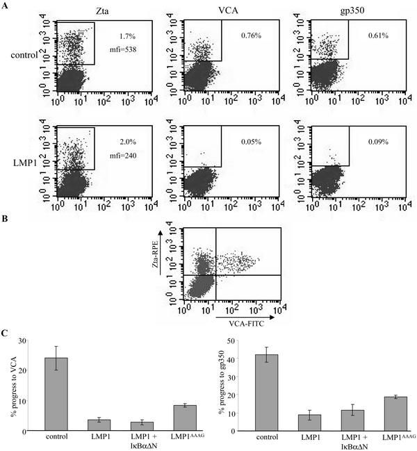
Epstein-Barr virus (EBV) is a potent growth-transforming agent of human B cells. It has previously been shown that viral latent membrane protein 1 (LMP1) is essential for EBV-induced transformation of normal B cells and contributes to maintenance of latency in vitro. Using the EBV-positive Burkitt's lymphoma line P3HR1-c16, which lacks LMP1 during latency and which can readily be activated into virus-productive lytic cycle, we found that LMP1 inhibits lytic cycle induction via the transcription factor NF-kappa B. In addition, LMP1 inhibits lytic cycle progress via two distinct NF-kappa B-independent mechanisms: one involving the cytosolic C-terminal activating regions and the other involving the transmembrane region of LMP1. These findings indicate that in B cells EBV self-limits its lytic cycle via three distinct LMP1-mediated mechanisms.


Figures

FIG. 1.
FIG. 1.
LMP1-mediated inhibition of EBV lytic cycle induction. (A) Zta expression in cells induced to lytic cycle with sodium butyrate. P3HR1-c16 cells were cultured and electroporated, using methods described previously (32), with 6 μg of plasmid rCD2-EGFP and either 3 μg of empty pSG5 vector (control) or 3 μg of the B95.8 LMP1 expression plasmid, pSG5-LMP1 (26). Electroporation was performed using a Bio-Rad Genepulser II electroporator set at 950 μF and 240 V. The cells were seeded in 4 ml of fresh medium and cultured for 48 h before induction to the lytic cycle by resuspension in fresh medium containing 3 mM sodium butyrate. The cells were cultured for a further 24 h, fixed, permeabilized, and stained for Zta expression as described previously (32) using 1 μg of murine monoclonal antibody BZ.1 (58) per ml, and red phycoerythrin (RPE)- conjugated anti-mouse immunoglobulin G. The cells were analyzed by flow cytometry (40,000 events) using a FACScalibur flow cytometer (Becton Dickinson Co., San Jose, Calif.). The left panel shows a dot plot of the data obtained for cells cotransfected with empty pSG5 vector; the right panel shows data for the cells cotransfected with LMP1 expression plasmid. (B) Mechanism of LMP1-mediated inhibition of EBV lytic cycle induction. The experiment described above was repeated in triplicate and extended to include cotransfections of 6 μg of rCD2-EGFP expression plasmid with 3 μg of pSG5-LMP1AAAG expression plasmid (6) or 3 μg of pSG5-LMP1 plus 0.2 μg of IκBαΔN expression plasmids. Lytic cycle induction was calculated as the percentage of EGFP-positive cells that expressed Zta. The histogram shown displays means and standard errors (indicated by the error bars). One-way ANOVA P values and pooled-variance 95% confidence intervals were calculated using Minitab software (Minitab Inc., Pennsylvania State University). (C) Western blot analysis of LMP1 expression. Protein extracts of the cotransfections from panel B were analyzed for LMP1 expression by sodium dodecyl sulfate-polyacrylamide gel electrophoresis and immunoblotting with murine monoclonal antibodies CS.1 to CS.4 (46) as described previously (17, 47). The positions of molecular size standards (in kilodaltons) are shown to the left of the gel.
FIG. 2.
FIG. 2.
Effect of basal NF-κB activity on EBV lytic cycle induction. (A) Effect of increasing IκBαΔN expression plasmid dose on lytic cycle induction. The induction and staining procedures described in the legend to Fig. 1A were performed on triplicate cotransfections of P3HR-c16 cells with 6 μg of rCD2-EGFP expression plasmid and 0-, 0.2-, and 2-μg doses of IκBαΔN expression plasmid. The total amount of DNA per cotransfection was kept constant by the addition of an appropriate amount of empty pSG5. Flow cytometry data were processed as described in the legend to Fig. 1B. (B) Effect of increasing IκBαΔN expression plasmid dose on basal NF-κB activity. The cotransfections for panel A were repeated to include 3 μg of the NF-κB luciferase reporter plasmid 3Enh.κB-ConALuc (3). The cells were assayed for luciferase activity 24 h later as described previously (37). The histogram displays means and standard errors (indicated by the error bars).
FIG. 3.
FIG. 3.
Effect of LMP1 on early EBV lytic cycle. (A) Effect of LMP1 on the expression of early lytic cycle antigens. P3HR1-c16 cells were cotransfected with 8 μg of p509 Zta expression plasmid and either 3 μg of empty pSG5 vector (control), 3 μg of LMP1 expression plasmid, 3 μg of LMP1 plus 0.2 μg of IκBαΔN expression plasmids, or 3 μg of LMP1AAAG expression plasmid, as described in the legend to Fig. 1. The total amount of DNA per cotransfection was kept constant by the addition of an appropriate amount of empty pSG5. After 24 h, the cells were probed for expression of early lytic cycle antigens by immunoblotting with EE serum as described previously (32). To confirm equal loads in the lanes, the blot was stripped and reprobed for actin using antiactin antibodies as described previously (47). (B) Expression of exogenous Zta and LMP1. The cotransfections and immunoblots described above were repeated in EBV-negative DG75 cells electroporated at 270 V. Immunoblots were probed with BZ.1 antibody to Zta, CS.1 to CS.4 antibodies to LMP1, or antiactin antibodies, as described previously (32, 47). The positions of molecular size markers (in kilodaltons) are shown to the left of the blots.
FIG. 4.
FIG. 4.
Effect of LMP1 on Zta expression. The cotransfections described in the legend to Fig. 3A were repeated in triplicate to include 4 μg of the luciferase reporter plasmid for Zta activity, pHEBO:Zp-wt-luc. (A) Flow cytometric analysis of Zta expression. After 24 h, samples of cells were fixed, permeabilized, and analyzed for Zta expression by flow cytometry, as described in the legend to Fig. 1. (B) Zta transcriptional activity in cotransfected cells. Samples of cells were assayed for luciferase activity as described previously (37). Cells cotransfected with empty pSG5 in place of the p509 Zta expression plasmid were included as a negative control. Both histograms shown display means and standard errors (indicated by the error bars).
FIG. 5.
FIG. 5.
Effect of LMP1 on progress to late EBV lytic cycle. The cotransfections described in the legend to Fig. 3A were repeated in triplicate. After 48 h, the cells were fixed, permeabilized, and stained for Zta as described in the legend to Fig. 1. (A) Expression of late lytic cycle antigens. VCA was detected using V3 murine ascitic fluid (diluted 1:200) (55) (provided by Gary Pearson, Georgetown University, Washington, D.C.), and gp350 was detected using 1 μg of murine monoclonal antibody 72A1 (25) per ml. Primary antibody staining was detected by subsequent staining with red phycoerythrin (RPE)-conjugated anti-mouse immunoglobulin G (IgG) F(ab′)2 fragments (DAKO) (diluted 1:50). Antibody incubations were for 30 min at 37°C. The cells were analyzed by flow cytometry (as described in the legend to Fig. 1). The top row shows dot plots of data for Zta, VCA, and gp350 staining from a representative cotransfection with empty pSG5 vector; the bottom row shows the same for a pSG5-LMP1 cotransfection. mfi, mean fluorescence intensity. (B) Expression of Zta in late lytic cycle. P3HR1-c16 cells were transfected with 8 μg of Zta expression plasmid, and 48 h later, the cells were fixed and permeabilized (32). The cells were stained for simultaneous expression of Zta and VCA by use of antibodies BZ.1 and V3, respectively (32). BZ.1 staining was detected with RPE-conjugated anti-mouse IgG1 monoclonal antibody, and V3 staining was detected with fluorescein isothiocyanate (FITC)-conjugated anti-mouse IgG2a monoclonal antibody. The cells were analyzed by two-color flow cytometry (as described in the legend to Fig. 1), and a dot plot of the data was obtained. (C) Effect of LMP1 on progress to late lytic cycle. The total flow cytometry data obtained in panel A were processed to represent lytic cycle progress. For a given cotransfection, the percentage of VCA- or gp350-positive cells was divided by the percentage of Zta-positive cells and multiplied by 100. The histograms shown display means and standard errors (indicated by the error bars).
FIG. 6.
FIG. 6.
The LMP1 transmembrane region impedes progress to late EBV lytic cycle. The experimental procedures described in the legend to Fig. 5A and C were repeated for triplicate cotransfections of 8 μg of Zta expression plasmid with either 3 μg of empty pEGFP-N1 vector, 3 μg of EGFP-LMP1, 3 μg of EGFP-stop195LMP1, or 3 μg of rCD2-EGFP expression plasmids. The histograms display means and standard errors (indicated by the error bars).

References

    1. Adamson, A. L., and S. Kenney. 2001. Epstein-Barr virus immediate-early protein BZLF1 is SUMO-1 modified and disrupts promyelocytic leukemia bodies. J. Virol. 75:2388-2399. - PMC - PubMed
    1. Adler, B., E. Schaadt, B. Kempkes, U. Zimber-Strobl, B. Baier, and G. W. Bornkamm. 2002. Control of Epstein-Barr virus reactivation by activated CD40 and viral latent membrane protein 1. Proc. Natl. Acad. Sci. USA 99:437-442. - PMC - PubMed
    1. Arenzana-Seisdedos, F., B. Fernandez, I. Dominguez, J. M. Jacque, D. Thomas, M. T. Diaz-Meco, J. Moscat, and J. L. Virelizier. 1993. Phosphatidylcholine hydrolysis activates NF-κB and increases human immunodeficiency virus replication in human monocytes and T lymphocytes. J. Virol. 67:6596-6604. - PMC - PubMed
    1. Berberich, I., G. L. Shu, and E. A. Clark. 1994. Cross-linking CD40 on B cells rapidly activates nuclear factor-kappa B. J. Immunol. 153:4357-4366. - PubMed
    1. Borza, C. M., and L. M. Hutt-Fletcher. 2002. Alternate replication in B cells and epithelial cells switches tropism of Epstein-Barr virus. Nat. Med. 8:594-599. - PubMed

Publication types

MeSH terms

Substances