. 2003 Apr 8;107(13):1816-21.
doi: 10.1161/01.CIR.0000058461.86339.7E. Epub 2003 Mar 17.

Arrhythmogenic substrate of the pulmonary veins assessed by high-resolution optical mapping

Affiliations

Arrhythmogenic substrate of the pulmonary veins assessed by high-resolution optical mapping

Rishi Arora et al. Circulation. .

Abstract

Background: It has recently been recognized that atrial fibrillation can originate from focal sources in the pulmonary veins (PVs). However, the mechanisms of focal atrial fibrillation have not been well characterized. We assessed the electrophysiological characteristics of the PVs using high-resolution optical mapping.

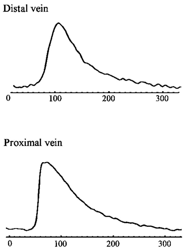
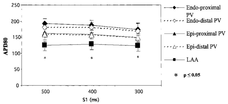
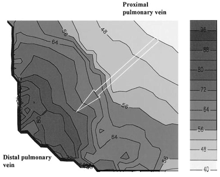
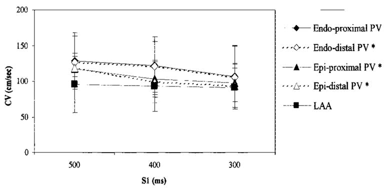
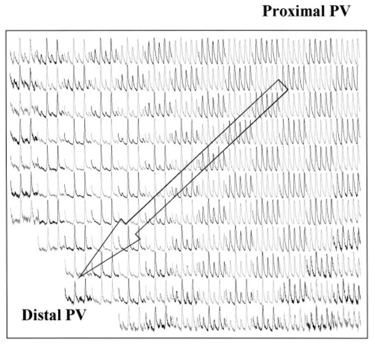
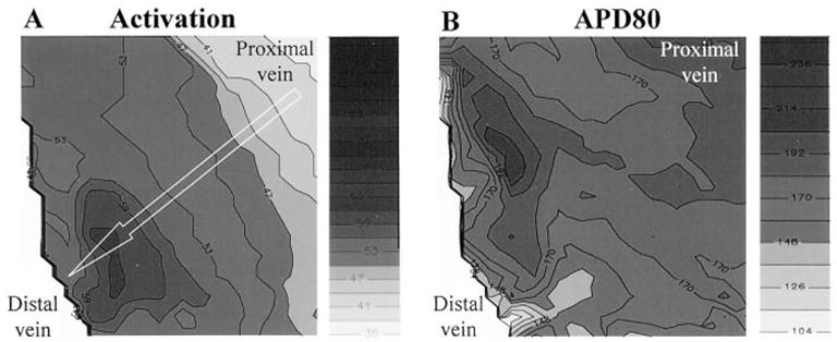
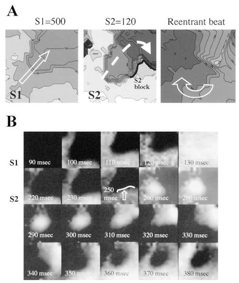
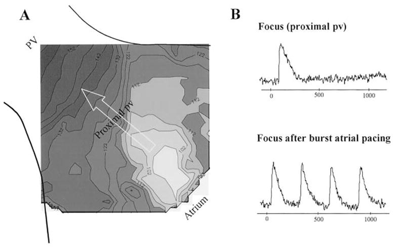
Methods and results: Coronary-perfused, isolated whole-atrial preparations from 33 normal dogs were studied. Programmed electrical stimulation was performed, and a 4-cm2 area of the PV underwent optical mapping of transmembrane voltage to obtain 256 simultaneous action potentials. Marked conduction slowing was seen at the proximal PV, compared with the rest of the vein, on both the epicardial (31.3+/-4.47 versus 90.2+/-20.7 cm/s, P=0.001) and endocardial (45.8+/-6.90 versus 67.6+/-10.4 cm/s, P=0.012) aspects. Pronounced repolarization heterogeneity was also noted, with action potential duration at 80% repolarization being longest at the PV endocardium. Nonsustained reentrant beats were induced with single extrastimuli, and the complete reentrant loop was visualized (cycle length, 155+/-30.3 ms); reentrant activity could be sustained with isoproterenol. Sustained focal discharge (cycle length, 330 to 1100 ms) was seen from the endocardial surface in the presence of isoproterenol; each focus was localized near the venous ostium.

Conclusions: The normal PV seems to have the necessary substrate to support reentry as well as focal activity. Although reentry occurred more distally in the vein, focal activity seemed to occur more proximally.


Figures

Figure 1
Figure 1
Typical optical action potentials (single pixels) obtained from proximal (bottom) and distal (top) aspects of vein. Action potential upstroke is significantly slower at distal vein than proximal vein.
Figure 2
Figure 2
Repolarization heterogeneity across PV and left atrium. APD80 at drive cycle lengths of 500, 400, and 300 ms are shown for proximal and distal PV for both endocardial (endo) and epicardial (epi) aspects and LAA. APD80 is longest at PV endocardium (endo>epi>LAA, *P≤0.05 at S1= 500 and 400 ms; at S1=300 ms, epi>LAA, endo>LAA, *P≤0.05).
Figure 3
Figure 3
Isochronal map of epicardial PV obtained during pacing at 500 ms. Scale is shown at right, with white being earliest activation and dark gray/black being latest activation. Pacing is occurring from left atrium (outside of field of view), which is at upper right of field of view. There is marked crowding of isochrones at junction of proximal vein and distal vein, signifying slow conduction in this region of PV. Arrow marks direction of wave-front propagation.
Figure 4
Figure 4
Decremental conduction in PV. Graph of conduction in PVs and left atrium at drive cycle lengths of 500, 400, and 300 ms. There is progressive slowing of conduction velocity, with a decrease in pacing cycle length, on both epicardial (epi) and endocardial (endo) aspects; *P≤0.05 at proximal and distal aspects of epicardial vein and at distal aspect of endocardial vein. No such conduction slowing is noted at LAA.
Figure 5
Figure 5
Optically derived action potentials during pacing at 250 ms, with 2:1 block in PV. Array demonstrates 4 action potentials per pixel in proximal vein at paced cycle length. In more distal portion of vein (lower left side of array), there are only 2 action potentials at half cycle length of pacing (2:1 block). Arrow marks direction of wave-front propagation.
Figure 6
Figure 6
Isochronal maps of activation and contour map of APD80 for same region of epicardial vein during same drive train. A, Isochronal map was obtained during atrial pacing at 500 ms. There is significant slowing of conduction at proximal vein, as shown by crowding of isochrones; arrow marks direction of wave-front propagation. B, APD80 for same impulse. Longest APD80 is noted in distal vein, 0.5 cm distal to region of slowest conduction in proximal vein.
Figure 7
Figure 7
Activation maps demonstrating reentry within PV (epicardial aspect) produced by a single extrastimulus. A, Left, With atrial pacing (S1=500 ms), there is slow conduction at proximal vein during impulse propagation, as indicated by crowding of isochrones in middle of field of view. Arrow marks direction of wave-front propagation. Middle and right, A premature extra-stimulus (S2=120) blocks at region of longest APD80 (region of block shown as thick black line) but continues to propagate through rest of region of slow conduction (dashed arrow) and then turns around to depolarize rest of proximal vein, which has since recovered (transparent arrow at right marks reentrant wave front). B, Time sequence of raw fluorescence signal from same beats as in A. White represents more positive voltage (ie, local depolarization), and dark blue represents more negative voltage (ie, resting). Sequences between final activation wave front of S1 and onset of wave front from S2 (diastolic period of field showing only resting voltage) have been omitted to conserve space. During activation of S1 (90 to 130 ms), a central area of slow conduction is seen. During activation of S2, activation of right side of wave front blocks (block shown as arrow and thick, black line) at area of prolonged APD80 (250-ms frame). Central area of slow conduction persists, with eventual rotation of wave front through previously blocked region (260 to 380 ms).
Figure 8
Figure 8
Isochronal map and representative action potentials during spontaneous focal activity observed during isoproterenol infusion. A, Isochronal map (format similar to that in previous figures) shows 1 beat from a spontaneous focus in proximal vein. Focus lies close to venous ostium (earliest activity at right lower corner of map) and lies 1.25 cm from zone of slow conduction in distal vein (crowded isochrones). Arrow marks direction of wave-front propagation. B, Representative optical action potentials recorded from focus (site of earliest activation). Top, Optical action potential from focus. Rate of focus on isoproterenol (10−6 mol/L) is 1100 ms. Bottom, With burst atrial pacing (not shown), rate of discharge of focus transiently increases to 300 ms (see text).

Similar articles

Cited by

References

    1. Chen SA, Chiang CE, Yang CJ, et al. Sustained atrial tachycardia in adult patients. Electrophysiological characteristics, pharmacological response, possible mechanisms, and effects of radiofrequency ablation. Circulation. 1994;90:1262–1278. - PubMed
    1. Haissaguerre M, Jais P, Shah DC, et al. Catheter ablation of chronic atrial fibrillation targeting the reinitiating triggers. J Cardiovasc Electrophysiol. 2000;11:2–10. - PubMed
    1. Chen SA, Hsieh MH, Tai CT, et al. Initiation of atrial fibrillation by ectopic beats originating from the pulmonary veins: electrophysiological characteristics, pharmacological responses, and effects of radiofrequency ablation. Circulation. 1999;100:1879–1886. - PubMed
    1. Wu J, Olgin J, Miller JM, et al. Mechanisms underlying the reentrant circuit of atrioventricular nodal reentrant tachycardia in isolated canine atrioventricular nodal preparation using optical mapping. Circ Res. 2001;88:1189–1195. - PubMed
    1. Verheule S, Wilson E, Arora R, et al. Tissue structure and connexin expression of canine pulmonary veins. Cardiovasc Res. 2002;55:727–738. - PubMed

Publication types