. 2003 Apr;23(8):2762-77.
doi: 10.1128/MCB.23.8.2762-2777.2003.

Molecular dissection of a yeast septin: distinct domains are required for septin interaction, localization, and function

Affiliations

Molecular dissection of a yeast septin: distinct domains are required for septin interaction, localization, and function

Antonio Casamayor et al. Mol Cell Biol. 2003 Apr.

Abstract

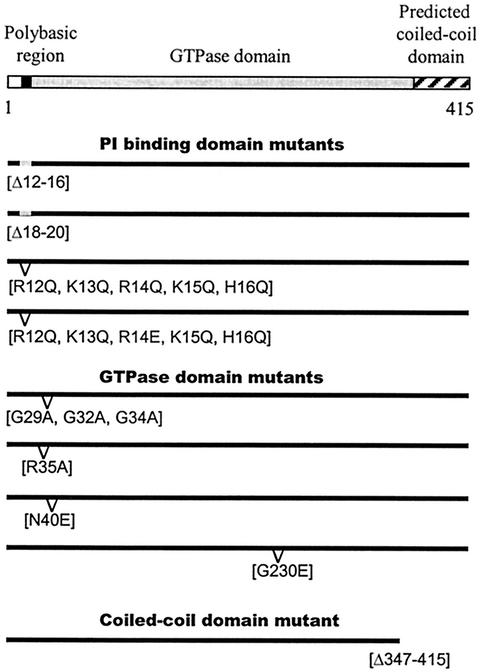
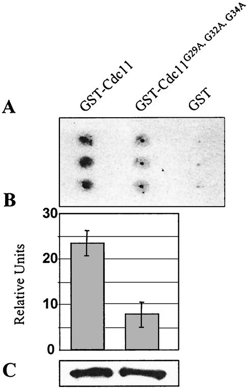
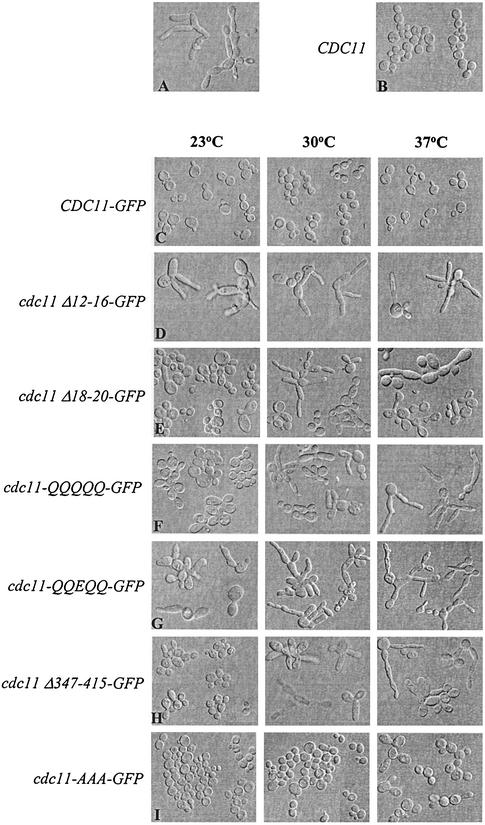
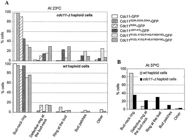
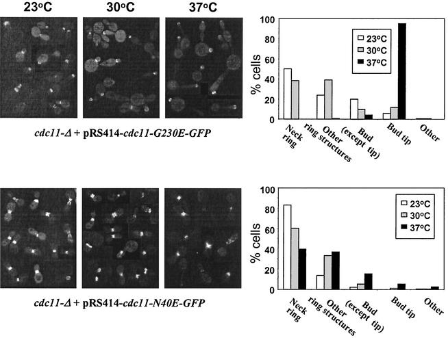
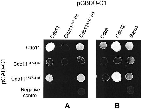
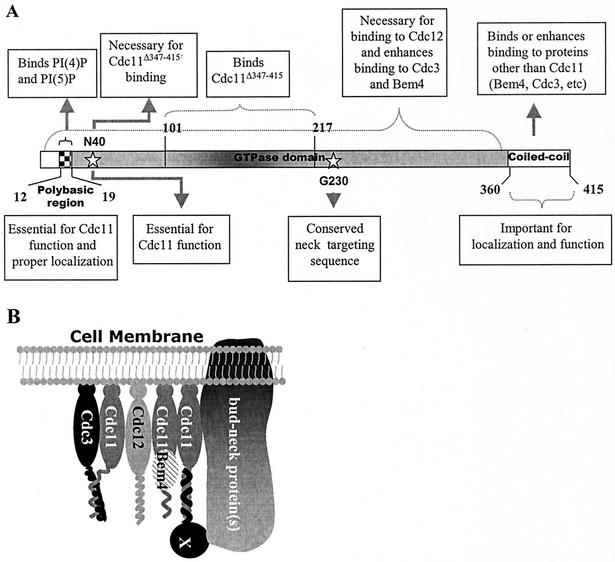
The septins are a family of cytoskeletal proteins present in animal and fungal cells. They were first identified for their essential role in cytokinesis, but more recently, they have been found to play an important role in many cellular processes, including bud site selection, chitin deposition, cell compartmentalization, and exocytosis. Septin proteins self-associate into filamentous structures that, in yeast cells, form a cortical ring at the mother bud neck. Members of the septin family share common structural domains: a GTPase domain in the central region of the protein, a stretch of basic residues at the amino terminus, and a predicted coiled-coil domain at the carboxy terminus. We have studied the role of each domain in the Saccharomyces cerevisiae septin Cdc11 and found that the three domains are responsible for distinct and sometimes overlapping functions. All three domains are important for proper localization and function in cytokinesis and morphogenesis. The basic region was found to bind the phosphoinositides phosphatidylinositol 4-phosphate and phosphatidylinositol 5-phosphate. The coiled-coil domain is important for interaction with Cdc3 and Bem4. The GTPase domain is involved in Cdc11-septin interaction and targeting to the mother bud neck. Surprisingly, GTP binding appears to be dispensable for Cdc11 function, localization, and lipid binding. Thus, we find that septins are multifunctional proteins with specific domains involved in distinct molecular interactions required for assembly, localization, and function within the cell.


Figures

FIG. 1.
FIG. 1.
Schematic representations of the cdc11 alleles used in this study. cdc11-Δ12-16 (Δ12-16) and cdc11-Δ18-20 (Δ18-20) contain deletions of five and three residues, respectively, located in the polybasic region. The cdc11-R12Q, K13Q, R14Q, K15Q, H16Q mutant (R12Q, K13Q, R14Q, K15Q, H16Q) has the positively charged residues deleted in cdc11-Δ12-16 replaced by the neutral glutamine (Q). cdc11-R12Q, K13Q, R14E, K15Q, H16Q (R12Q, K13Q, R14E, K15Q, H16Q) is similar to the previous mutant but has a glutamic acid (E) in position 14. cdc11-G29A, G32A, G34A (G29A, G32A, G34A) has been mutated in its nucleotide-binding P loop by replacing three glycine residues with alanine. The cdc11-R35A (R35A), cdc1-G230E (G230E), and cdc11-N40E (N40E) alleles have the indicated point mutations, and the coiled-coil domain of Cdc11 has been removed in the cdc11-Δ347-415 (Δ347-415) mutant.
FIG. 2.
FIG. 2.
Effect of the mutation Cdc11G29A, G32A, G34A on GTP binding. Equal amounts of purified GST-Cdc11 and GST-Cdc11G29A, G32A, G34A proteins were incubated in triplicate in the presence of [α-32P]GTP before being filtered onto a nitrocellulose membrane. The [α-32P]GTP present on the membrane after washes was detected by autoradiography (A), and the intensity of the spots was quantified (B). The quantity of protein present in each sample was determined by immunodetection, using an anti-GST antibody (C).
FIG. 3.
FIG. 3.
Effect of expression of different cdc11 alleles in cdc11-Δ cells. Strains were cultured overnight at 23°C, diluted, and then shifted to 23, 30, or 37°C. After 7 h of growth at each temperature, log-phase cells were fixed and the cellular morphology was observed. A representative composite is shown in each case. (A) cdc11-Δ cells (strain YAC660) were cultured for 3 days at 23°C due to its low growth rate. (B) Strain cdc11-Δ expressing Cdc11 from the centromeric vector pRS414 under control of its own promoter (YAC685) was grown at 30°C. (C) cdc11-Δ cells expressing Cdc11-GFP from pRS414 under the control of its own promoter (YAC731); (D) cdc11-Δ cells carrying pRS414-cdc11 Δ12-16-GFP (YAC780); (E) cdc11-Δ cells expressing cdc11-Δ18-20-GFP (YAC728); (F) cdc11-Δ cells carrying pRS414-cdc11-R12Q, K13Q, R14Q, K15Q, H16Q-GFP (YAC804); (G) cdc11-Δ cells expressing cdc11-R12Q, K13Q, R14E, K15Q, H16Q-GFP (YAC808); (H) cdc11-Δ cells expressing cdc11Δ-347-415-GFP (YAC793); (I) cdc11-Δ cells carrying pRS414-cdc11-G29A, G32A, G34A-GFP (YAC715); (J) cdc11-Δ cells carrying pRS414-cdc11-G230E-GFP (YAC704); (K) cdc11-Δ cells expressing cdc11-N40E-GFP (YAC881).
FIG. 3.
FIG. 3.
Effect of expression of different cdc11 alleles in cdc11-Δ cells. Strains were cultured overnight at 23°C, diluted, and then shifted to 23, 30, or 37°C. After 7 h of growth at each temperature, log-phase cells were fixed and the cellular morphology was observed. A representative composite is shown in each case. (A) cdc11-Δ cells (strain YAC660) were cultured for 3 days at 23°C due to its low growth rate. (B) Strain cdc11-Δ expressing Cdc11 from the centromeric vector pRS414 under control of its own promoter (YAC685) was grown at 30°C. (C) cdc11-Δ cells expressing Cdc11-GFP from pRS414 under the control of its own promoter (YAC731); (D) cdc11-Δ cells carrying pRS414-cdc11 Δ12-16-GFP (YAC780); (E) cdc11-Δ cells expressing cdc11-Δ18-20-GFP (YAC728); (F) cdc11-Δ cells carrying pRS414-cdc11-R12Q, K13Q, R14Q, K15Q, H16Q-GFP (YAC804); (G) cdc11-Δ cells expressing cdc11-R12Q, K13Q, R14E, K15Q, H16Q-GFP (YAC808); (H) cdc11-Δ cells expressing cdc11Δ-347-415-GFP (YAC793); (I) cdc11-Δ cells carrying pRS414-cdc11-G29A, G32A, G34A-GFP (YAC715); (J) cdc11-Δ cells carrying pRS414-cdc11-G230E-GFP (YAC704); (K) cdc11-Δ cells expressing cdc11-N40E-GFP (YAC881).
FIG. 4.
FIG. 4.
Analysis of the phosphoinositide-binding properties of yeast septins. The ability of the indicated yeast GST-septins to bind to different lipids was analyzed using a protein lipid overlay assay (11). Abbreviations: S 1-P, sphingosine 1-phosphate; PA, phosphatidic acid; PS, phosphatidylserine; LPA, lysophosphatidic acid; LPC, lysophosphocholine; PE, phosphatidylethanolamine; PC, phosphatidylcholine.
FIG. 5.
FIG. 5.
Lipid-binding properties of GST-Cdc11. (A) Wild-type and mutant GST-Cdc11 fusion proteins were tested for their lipid-binding properties as described in the legend to Fig. 4. (B) GST-Cdc11 fusion protein was loaded with GDP and γ-S-labeled GTP before incubation with the lipid-containing membrane. (C) Alignment of the polybasic region located 10 residues amino terminal with respect to the P loop in the mammalian H5 protein and the seven septins encoded in the yeast genome. The one-letter code for amino acids is used. Basic residues are shown in bold type. Asterisks mark the five positively charged amino acids replaced in the GST-Cdc11R12Q, K13Q, R14Q, K15Q, H16Q and GST-Cdc11R12Q, K13Q, R14E, K15Q, H16Q mutants. Note that the location of the basic residues in Spr3 is further from the P loop than for the other septins. (D) Protein lipid overlay assay using the Cdc11 fusion mutants where the five basic residues in positions 12 to 16 have been replaced as indicated. (E) Pull-down assay. The Western blots were treated with an anti-GST antibody. The bands in lanes 1 and 2 show the input protein, and the bands in lanes 3 and 4 show protein associated with the PI(4)P agarose beads. Lanes 1 and 3 contain wild-type GST-Cdc11, and lanes 2 and 4 contain GST-Cdc11R12Q, K13Q, R14E, K15Q, H16Q.
FIG. 6.
FIG. 6.
Quantification of budded yeast cells with undetectable Cdc11-GFP in mutants lacking PI(4)P. Strains SEY6210 (wild type [wt]), AAY102 (stt4-4), AAY104 (pik1-83), and AAY105 (stt4-4 pik1-83) (2) were transformed with a centromeric plasmid expressing Cdc11-GFP under the CDC11 promoter. Log-phase cultures of the corresponding strains were grown at the indicated temperature for 3 h before fixation. The number of budded cells displaying Cdc11-GFP signal was quantified in at least 250 cells. The percentage of budded cells in which Cdc11-GFP staining was not observed is plotted on the y axis.
FIG. 7.
FIG. 7.
Subcellular localization of GFP-tagged cdc11 alleles. cdc11Δ (A to E) and CDC11 (wild type) (F to J) cells were transformed with centromeric plasmids expressing Cdc11-GFP (A and F), Cdc11Δ347-415-GFP (B and G), Cdc11-R12Q, K13Q, R14E, K15Q, H16Q-GFP (C and H), Cdc11R12Q, K13Q, R14Q, K15Q, H16Q-GFP (D and I), or Cdc11G29A, G32A, G34A-GFP (E and J). Cells were grown overnight at 23°C, diluted into fresh SC medium lacking Trp, grown until mid-exponential phase at the same temperature, and fixed. Different localization patterns are indicated by the arrows.
FIG. 8.
FIG. 8.
Quantification of the subcellular localization of different alleles of Cdc11-GFP in cdc11-Δ and CDC11 (wild-type [wt]) cells. (A) The localization of different alleles of Cdc11-GFP cells, treated as indicated in the legend to Fig. 7, was examined. More than 200 budded cells were observed in each category, and the results are presented as a percentage of the total number of budded cells. (B) Localization of the Cdc11R35A-GFP allele at 37°C in both wild-type and cdc11-Δ mutant cells.
FIG. 9.
FIG. 9.
Subcellular distribution of alleles of GFP-tagged Cdc11 with point mutations in a cdc11-Δ background at different temperatures. cdc11-Δ cells expressing either Cdc11G230E-GFP or Cdc11N40E-GFP from a centromeric plasmid were grown overnight, diluted in fresh SC medium lacking Trp, and grown until mid-exponential phase for 7 h at the indicated temperatures. The cellular localization of Cdc11G230E-GFP and Cdc11N40E-GFP was examined in more than 200 budded cells. Examples of localization of both alleles are shown in the micrographs on the left, while quantification of the localization of alleles for the indicated categories at each temperature are shown in the graphs on the right.
FIG. 10.
FIG. 10.
Two-hybrid interactions between Cdc11 and other yeast proteins. Six-microliter samples of log-phase cultures at similar cell densities were spotted onto plates containing SC medium minus Ura, Leu, and His. The yeast cells carry two plasmids: (i) the pGAD-C1 plasmid (marked with LEU2) harboring fragments of DNA encoding either the complete coding region of CDC11 (Cdc11), the coiled-coil domain of Cdc11 (Cdc11347-415), Cdc11 lacking its coiled-coil domain (Cdc11-Δ347-415), or the plasmid without any insert as a negative control, and (ii) the pGBDU-C1 plasmid (marked with URA3), carrying the already described fragments of CDC11 (A) or the full coding region of CDC3, CDC12, or BEM4. An interaction between different fragments leads to expression of the reporter gene HIS3 that enables growth on the plate.
FIG. 11.
FIG. 11.
Mapping the region of Cdc11 required for interaction with itself. (A) Yeast strains harboring the pGAD-C1 plasmid (marked with LEU2) expressing wild-type Cdc11 or Cdc11Δ347-415 or lacking an insert and the pGBDU-C1 plasmid (marked with URA3) carrying DNA encoding the indicated fragments of CDC11 are shown. Yeast cells were grown on SC medium minus Ura, Leu, and His; only cells carrying fragments of Cdc11 capable of interacting with Cdc11 can grow on this medium. (B) Yeast strains harboring the pGBDU-C1 plasmid expressing Cdc11Δ347-415 or Cdc11Δ347-415, N40E and pGAD-C1 plasmid expressing Cdc11, Cdc11347-415, or Cdc11Δ347-415 were grown on SC medium minus Ura, Leu, and His. Six microliters of each yeast liquid culture carrying the indicated constructs were spotted onto a plate containing the same medium. (C) Schematic representation of the truncated Cdc11 proteins used in the two-hybrid analyses. The complete coiled-coil domain of Cdc11 is included in the fragment Cdc11347-415. The fragments capable of interacting with Cdc11 (as detected by two-hybrid analysis) are shown in black boxes.
FIG. 12.
FIG. 12.
A model for Cdc11 function. (A) Summary of the different regions of Cdc11 required for its interactions and activity. (B) A model for how Cdc11 associates with other proteins at the bud neck. A putative protein(s) that interacts with Cdc11 through the coiled-coil domain and with other proteins anchored to the bud neck is indicated by the X.

References

    1. Audhya, A., and S. D. Emr. 2002. Stt4 PI 4-kinase localizes to the plasma membrane and functions in the Pkc1-mediated MAP kinase cascade. Dev. Cell 2:593-605. - PubMed
    1. Audhya, A., M. Foti, and S. D. Emr. 2000. Distinct roles for the yeast phosphatidylinositol 4-kinases, Stt4p and Pik1p, in secretion, cell growth, and organelle membrane dynamics. Mol. Biol. Cell 11:2673-2689. - PMC - PubMed
    1. Barral, Y., V. Mermall, M. S. Mooseker, and M. Snyder. 2000. Compartmentalization of the cell cortex by septins is required for maintenance of cell polarity in yeast. Mol. Cell 5:841-851. - PubMed
    1. Barral, Y., M. Parra, S. Bidlingmaier, and M. Snyder. 1999. Nim1-related kinases coordinate cell cycle progression with the organization of the peripheral cytoskeleton in yeast. Genes Dev. 13:176-187. - PMC - PubMed
    1. Byers, B., and L. Goetsch. 1976. A highly ordered ring of membrane-associated filaments in budding yeast. J. Cell Biol. 69:717-721. - PMC - PubMed

Publication types

MeSH terms