. 2003 Apr;23(8):2871-82.
doi: 10.1128/MCB.23.8.2871-2882.2003.

c-Jun NH(2)-terminal kinase is essential for the regulation of AP-1 by tumor necrosis factor

Affiliations

c-Jun NH(2)-terminal kinase is essential for the regulation of AP-1 by tumor necrosis factor

Juan-Jose Ventura et al. Mol Cell Biol. 2003 Apr.

Abstract

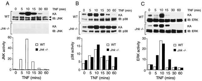
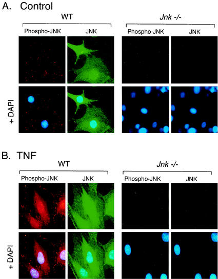
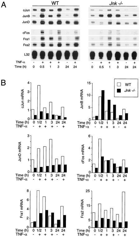
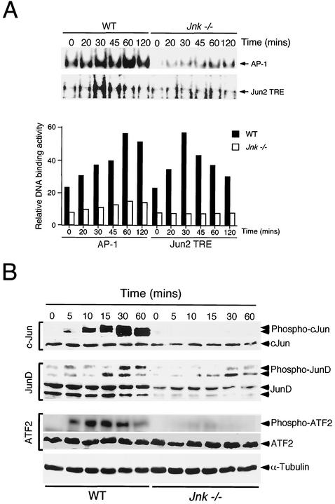
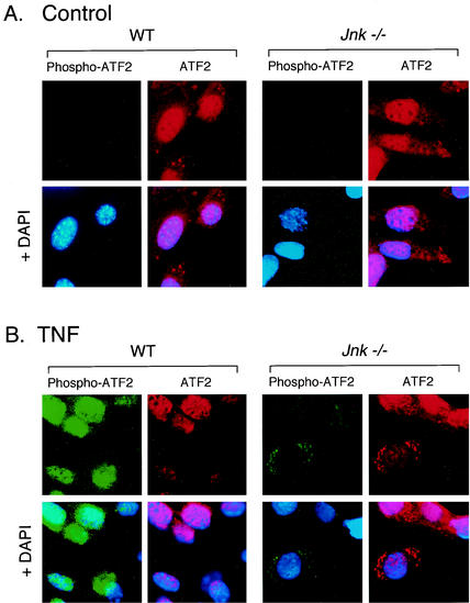
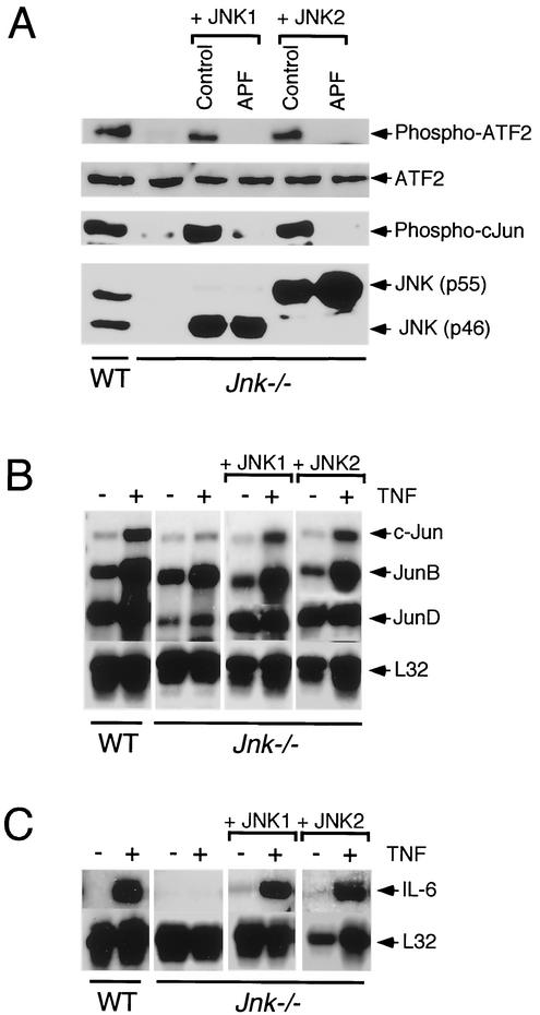
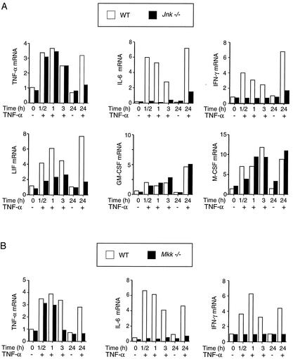
The c-Jun NH(2)-terminal kinase (JNK) is activated by the cytokine tumor necrosis factor (TNF). This pathway is implicated in the regulation of AP-1-dependent gene expression by TNF. To examine the role of the JNK signaling pathway, we compared the effects of TNF on wild-type and Jnk1(-/-) Jnk2(-/-) murine embryo fibroblasts. We show that JNK is required for the normal regulation of AP-1 by TNF. The JNK-deficient cells exhibited decreased expression of c-Jun, JunD, c-Fos, Fra1, and Fra2; decreased phosphorylation of c-Jun and JunD; and decreased AP-1 DNA binding activity. The JNK-deficient cells also exhibited defects in the regulation of the AP-1-related transcription factor ATF2. These changes were associated with marked defects in TNF-regulated gene expression. The JNK signal transduction pathway is therefore essential for AP-1 transcription factor regulation in cells exposed to TNF.


Figures

FIG. 1.
FIG. 1.
Effect of TNF on the activation of MAPK in WT and Jnk−/− fibroblasts. The activities of JNK (A), p38 MAPK (B), and ERK (C) in cells treated with TNF-α (10 ng/ml) for different times were measured in an immunocomplex protein kinase assay (KA). The amount of MAPK was examined by immunoblot (IB) analysis. The substrate phosphorylation in the kinase assays was quantitated using a PhosphorImager and is presented graphically in relative units. The data shown were derived from a single experiment and are representative of three independent experiments.
FIG. 2.
FIG. 2.
Comparison of JNK expression and activation in WT and Jnk−/− fibroblasts. JNK and activated JNK (Phospho-JNK) were examined by immunofluorescence microscopy. The cells were treated without (A) and with (B) TNF-α (10 ng/ml for 15 min), fixed, and stained with antibodies to JNK (FITC [green]) and phospho-JNK (Texas red [red]). DNA was stained with DAPI (blue). The cells were imaged by conventional fluorescence microscopy.
FIG. 3.
FIG. 3.
AP-1 transcription factor expression in WT and Jnk−/− fibroblasts. WT and Jnk−/− cells were treated without (−) and with (+) TNF-α (10 ng/ml). The cells were harvested after different times, and total RNA was isolated. The c-Jun, JunB, JunD, c-Fos, Fra1, Fra2, and ribosomal protein L32 mRNAs were examined in an RNase protection assay. The mRNAs were detected by autoradiography (A) and quantitated by PhosphorImager analysis (B). The normalized AP-1 mRNA/L32 mRNA ratio is presented. The data shown are representative of data obtained in three independent experiments.
FIG. 4.
FIG. 4.
AP-1 transcription factors in WT and Jnk−/− fibroblasts. (A) WT and Jnk−/− cells were treated without and with TNF-α (10 ng/ml). Nuclear extracts were prepared at different times and used to examine DNA binding activity with an electrophoretic mobility shift assay using probes containing a consensus AP-1 site (TRE) and a nonconsensus site (Jun2 TRE). The DNA binding activity was detected by autoradiography (upper panel) and was quantitated by PhosphorImager analysis (lower panel). (B) WT and Jnk−/− cells were treated without and with TNF-α (10 ng/ml). The cells were harvested after different times. Cell lysates were examined by immunoblot analysis by probing with antibodies to ATF2, c-Jun, and JunD. The phosphorylation of these transcription factors on sites that are phosphorylated by JNK was examined using phospho-specific antibodies. Blots were probed with an antibody to α-tubulin to monitor protein loading on each lane of the gel.
FIG. 5.
FIG. 5.
Effect of JNK deficiency on phosphorylation and subcellular location of ATF2. ATF2 and phospho-ATF2 were examined by immunofluorescence microscopy. Fibroblasts were treated without (A) and with (B) TNF-α (10 ng/ml for 15 min), fixed, and stained with antibodies to ATF2 (Texas red [red]) and phospho-ATF2 (FITC [green]). DNA was stained with DAPI (blue). The cells were imaged by conventional fluorescence microscopy.
FIG. 6.
FIG. 6.
Phosphorylation of ATF2 and c-Jun is restored in Jnk−/− fibroblasts following ectopic expression of JNK1 or JNK2. (A) Complementation analysis of Jnk−/− cells by ectopic expression of JNK1 or JNK2. Jnk−/− cells were transduced with retroviruses that expressed WT JNK1 or JNK2. Studies were also performed using retroviruses expressing kinase-inactive JNK1 or JNK2 (APF, replacement of the tripeptide dual phosphorylation motif Thr-Pro-Tyr with Ala-Pro-Phe). The WT cells, Jnk−/− cells, and the four complemented Jnk−/− cell populations were treated with TNF (15 min) and harvested to prepare lysates that were probed with antibodies to JNK, ATF2, phospho-ATF2, and phospho-c-Jun. (B and C) WT cells, Jnk−/− cells, and Jnk−/− cells complemented with JNK1 or JNK2 were treated without (−) and with (+) TNF-α (10 ng/ml for 60 min). Total RNA (5 μg) was isolated and examined using an RNase protection assay to measure the expression of c-Jun, JunB, and JunD (B) and IL-6 mRNA (C). Control assays were performed to measure the expression of the ribosomal protein L32.
FIG. 7.
FIG. 7.
ATF2 transcription activity in WT and Jnk−/− fibroblasts. ATF2 activity was examined in a luciferase reporter assay using the activation domain of ATF2 fused to the GAL4 DNA binding domain (14). The effect of replacement of the sites of activating phosphorylation (Thr-69 and Thr-71) with Ala was investigated (GAL4/ATF2A). The relative firefly luciferase activity was measured and was normalized to the amount of activity detected for a cotransfected control Renilla luciferase reporter plasmid. (A) The effect of coexpression of MEK kinase 1 (MEKK1), MKK6 (Ser208Glu/Thr-211Glu), and mixed-lineage kinase 3 (MLK3) was examined. (B) Complementation analysis of Jnk−/− cells by ectopic expression of JNK1 or JNK2. The cells were cotransfected with an expression vector for JNK1 or JNK2. Control studies were performed by cotransfecting the empty expression plasmid.
FIG. 8.
FIG. 8.
Role of JNK in TNF-stimulated cytokine expression. (A) WT and Jnk−/− fibroblasts were starved for 3 h and then treated with TNF-α (10 ng/ml). Total RNA was purified, and 5 μg was used for RNase protection assays to measure the amount of TNF-α, IL-6, IFN-γ, LIF, GM-CSF, M-CSF, and ribosomal protein L32 mRNA. The ratio of cytokine mRNA/L32 mRNA was calculated and is presented as the relative amount of cytokine expression. The data shown are representative of three independent experiments. (B) RNase protection assays were used to measure the amount of TNF-α, IL-6, and IFN-γ mRNA expressed by WT and Mkk−/− cells.

References

    1. Andrews, N. C., and D. V. Faller. 1991. A rapid micropreparation technique for extraction of DNA-binding proteins from limiting numbers of mammalian cells. Nucleic Acids Res. 19:2499.. - PMC - PubMed
    1. Baud, V., and M. Karin. 2001. Signal transduction by tumor necrosis factor and its relatives. Trends Cell Biol. 11:372-377. - PubMed
    1. Bennett, B. L., D. T. Sasaki, B. W. Murray, E. C. O'Leary, S. T. Sakata, W. Xu, J. C. Leisten, A. Motiwala, S. Pierce, Y. Satoh, S. S. Bhagwat, A. M. Manning, and D. W. Anderson. 2001. SP600125, an anthrapyrazolone inhibitor of Jun N-terminal kinase. Proc. Natl. Acad. Sci. USA 98:13681-13686. - PMC - PubMed
    1. Beyaert, R., A. Cuenda, W. Vanden Berghe, S. Plaisance, J. C. Lee, G. Haegeman, P. Cohen, and W. Fiers. 1996. The p38/RK mitogen-activated protein kinase pathway regulates interleukin-6 synthesis response to tumor necrosis factor. EMBO J. 15:1914-1923. - PMC - PubMed
    1. Brenner, D. A., M. O'Hara, P. Angel, M. Chojkier, and M. Karin. 1989. Prolonged activation of jun and collagenase genes by tumour necrosis factor-alpha. Nature 337:661-663. - PubMed

Publication types

MeSH terms

Substances