. 2003 Apr;23(8):2914-26.
doi: 10.1128/MCB.23.8.2914-2926.2003.

The carboxy terminus of the small subunit of TFIIE regulates the transition from transcription initiation to elongation by RNA polymerase II

Affiliations

The carboxy terminus of the small subunit of TFIIE regulates the transition from transcription initiation to elongation by RNA polymerase II

Tomomichi Watanabe et al. Mol Cell Biol. 2003 Apr.

Abstract

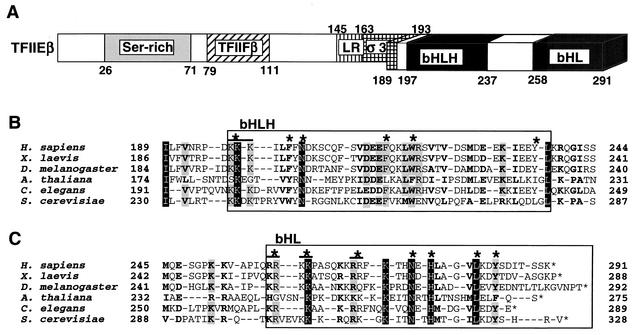
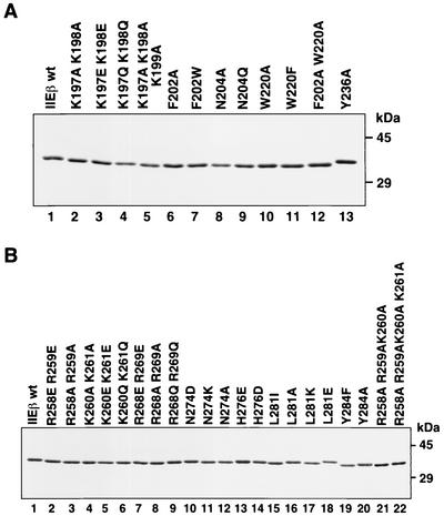
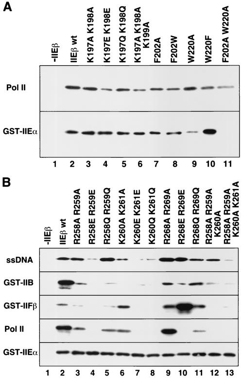
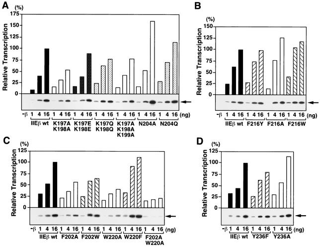
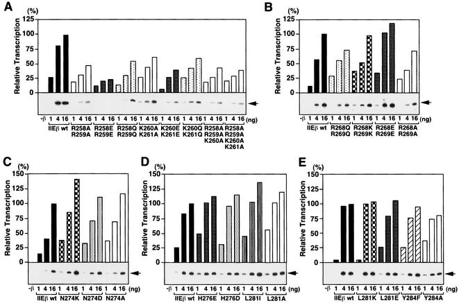
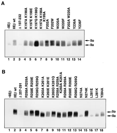
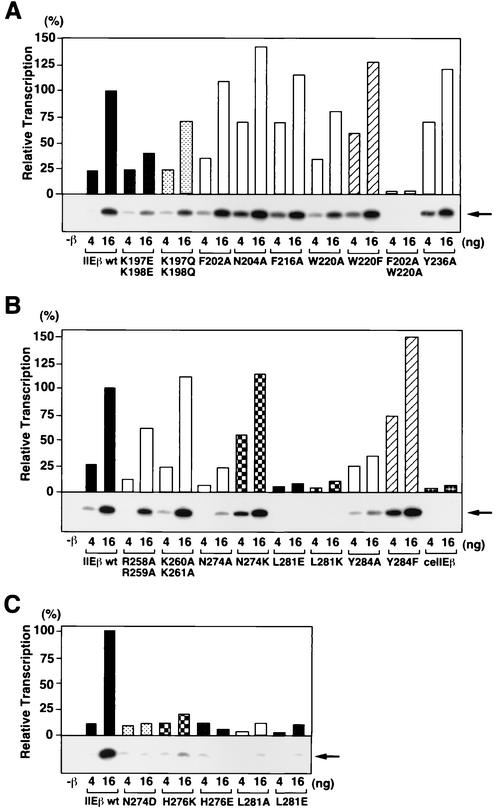
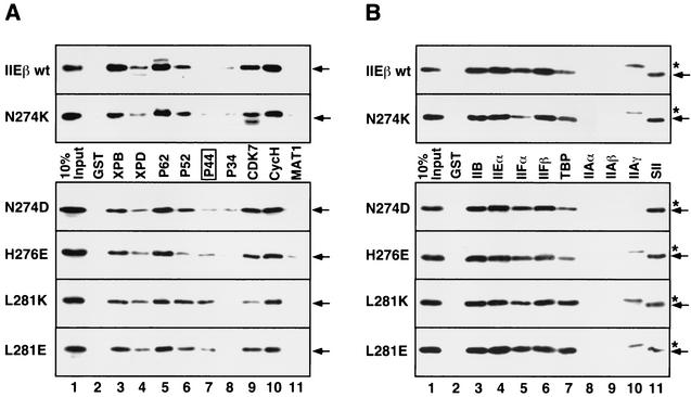
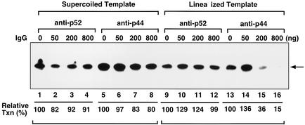
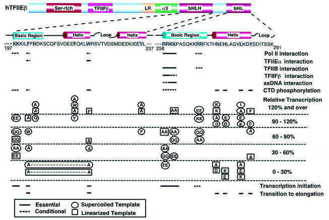
The general transcription factor TFIIE plays essential roles in both transcription initiation and the transition from initiation to elongation. Previously, we systematically deleted the structural motifs and characteristic sequences of the small subunit of human TFIIE (hTFIIE beta) to map its functional regions. Here we introduced point mutations into two regions located near the carboxy terminus of hTFIIE beta and identified the functionally essential amino acid residues that bind to RNA polymerase II (Pol II), the general transcription factors, and single-stranded DNA. Although most residues identified were essential for transcription initiation, use of an in vitro transcription assay with a linearized template revealed that several residues in the carboxy-terminal helix-loop region are crucially involved in the transition stage. Mutations in these residues also affected the ability of hTFIIE beta to stimulate TFIIH-mediated phosphorylation of the carboxy-terminal heptapeptide repeats of the largest subunit of Pol II. Furthermore, these mutations conspicuously augmented the binding of hTFIIE beta to the p44 subunit of TFIIH. The antibody study indicated that they thus altered the conformation of one side of TFIIH, consisting of p44, XPD, and Cdk-activating kinase subunits, that is essential for the transition stage. This is an important clue for elucidating the molecular mechanisms involved in the transition stage.


Figures

FIG. 1.
FIG. 1.
Structural features of hTFIIEβ. (A) Schematic diagram of the structural motifs and characteristic sequences of hTFIIEβ. Included residues are shown. Ser-rich, serine-rich sequence; TFIIFβ, region similar to the dsDNA-binding region of TFIIFβ (RAP30); LR, leucine repeat motif; σ3, region similar to the bacterial σ factor subdomain 3; bHLH, bHLH motif; bHL, a bHLH motif-like sequence that lacks the second helix. The C-terminal region (residues 189 to 291) containing bHLH and bHL has been drawn stereographically. (B) Sequence alignment of the bHLH region of hTFIIEβ with that of TFIIβ from the following species (sources of sequence data are in parentheses): Homo sapiens (48), Xenopus laevis (27), Drosophila melanogaster (54), Arabidopsis thaliana (GenBank accession no. CAB79101), C. elegans (56), and S. cerevisiae (7). Sequences corresponding to the bHLH motif are boxed. Asterisks above sequences, mutated residues. A double mutant is indicated by overlining and an asterisk. (C) Sequence alignment of the bHL region of hTFIIEβ with those of other species. Sequences corresponding to the bHL region are boxed. Mutated residues and double mutants are indicated as in panel B. Completely identical residues are shaded in black, and residues identical in five species are shaded in gray. Conserved similar residues are in boldface. Identical and similar amino acids were identified as described previously (27, 48).
FIG. 2.
FIG. 2.
Purified hTFIIEβ proteins with point mutations in the bHLH and bHL regions. (A) SDS-PAGE of the purified hTFIIEβ proteins with point mutations in the bHLH region. Histidine-tagged point mutants were expressed in E. coli BL21(DE3)pLysS and purified, and 400 ng of each was subjected to SDS-PAGE (12% acrylamide) and stained with Coomassie blue. Lane 1, wild-type hTFIIEβ; lanes 2 to 13, mutant proteins. The sizes of the molecular weight markers are indicated on the right. Mutated residues are indicated at the top of each lane. (B) SDS-PAGE of the purified hTFIIEβ proteins with point mutations in the bHL region. Expression, purification, and SDS-PAGE were performed as described for panel A. Lane 1, wild-type hTFIIEβ; lanes 2 to 20, mutant proteins.
FIG. 3.
FIG. 3.
Ability of the hTFIIEβ mutants to bind to ssDNA, general transcription factors, and Pol II. (A) Binding of hTFIIEβ proteins with point mutations in the bHLH region. Mutant hTFIIEβ proteins were mixed with Pol II (top) or GST-hTFIIEα (GST-IIEα; bottom) bound to protein G-Sepharose beads (Pol II) or glutathione-Sepharose resin (GST-IIEα). The mixtures were rotated for 4 h at 4°C, washed, and subjected to SDS-PAGE. Bound mutants were detected by Western blotting with anti-hTFIIEβ antisera. Lane 1, no hTFIIEβ was added (−IIEβ); lane 2, wild-type hTFIIEβ (IIEβ wt); lanes 3 to 11, point mutant proteins. Mutated residues are indicated at the top of each lane. (B) Binding of hTFIIEβ proteins with point mutations in the bHL region. Binding assays were performed as in panel A. ssDNA, GST-TFIIB (GST-IIB), GST-TFIIFβ (GST-IIFβ), Pol II, and GST-hTFIIEα (GST-IIEα) bound to agarose (ssDNA), glutathione-Sepharose (general transcription factors), or protein G-Sepharose beads (Pol II). Lane 1, no hTFIIEβ (−IIEβ); lane 2, wild-type hTFIIEβ; lanes 3 to 13, point mutant proteins. Mutated residues are indicated at the top of each lane.
FIG. 4.
FIG. 4.
Effect of bHLH point mutations on the basal transcription activity of hTFIIEβ with a supercoiled template. In vitro transcription assays with a supercoiled template were carried out with increasing amounts (1, 4, and 16 ng) of wild-type hTFIIEβ (IIEβ wt) or hTFIIEβ proteins with point mutations in the bHLH region. After the transcription reaction, radiolabeled transcripts were subjected to urea-PAGE and detected by autoradiography (bottom of each graph). Each transcript was quantified by a Fuji BAS2500 Bio-Imaging analyzer. Relative transcription activities (bars) of the mutant hTFIIEβ proteins were calculated by defining the transcription activity of 16 ng of wild-type hTFIIEβ as 100%. Mutated residues are indicated at the bottom of each panel. As a control, transcription was carried out without the hTFIIEβ protein (−β). Arrows (right side of each panel), positions of the 390-nt transcripts.
FIG. 5.
FIG. 5.
Effect of bHL point mutations on the basal transcription activity of hTFIIEβ with a supercoiled template. In vitro transcription assays were performed as described for Fig. 4 with wild-type hTFIIEβ (IIEβ wt) or hTFIIEβ proteins with mutations in the bHL region. Arrows are as defined for Fig. 4.
FIG. 6.
FIG. 6.
Effects of bHLH and bHL point mutations on TFIIH-mediated CTD phosphorylation. (A) bHLH point mutations. Kinase assays were performed under the conditions of active initiation complex formation. Lane 1, no hTFIIEβ; lane 2, wild-type (wt) hTFIIEβ; lane 3, hTFIIEβ bHLH deletion mutant; lanes 4 to 14, hTFIIEβ with point mutations in the bHLH region. The mutated residues are indicated at the top of each lane. The phosphorylation of the largest subunit of Pol II was analyzed on a 5.5% acrylamide-SDS gel and detected by autoradiography. Arrows, phosphorylated form of the largest subunit of Pol II (IIo) and the unphosphorylated form (IIa). (B) bHL point mutations. Kinase assays were performed as described for panel A. Lane 1, no hTFIIEβ; lane 2, wild-type hTFIIEβ; lane 3, hTFIIEβ bHL deletion mutant; lanes 4 to 18, hTFIIEβ with point mutations in the bHL region. Arrows, IIo and IIa.
FIG. 7.
FIG. 7.
Effects of bHLH and bHL point mutations on basal transcription activity with a linearized template. (A) bHLH point mutations. In vitro transcription assays were performed as described for Fig. 4 except with a linearized template. Increasing amounts (4 and 16 ng) of wild-type hTFIIEβ (IIEβ wt) and hTFIIEβ proteins with mutations in the bHLH region were tested. After the transcription reaction, the radiolabeled transcripts were subjected to urea-PAGE and detected by autoradiography (below graph). The transcripts were quantified with a Fuji BAS2500 Bio-Imaging analyzer. Relative transcription activities (bars) of the mutant hTFIIEβ proteins were calculated by defining the transcription activity of 16 ng of wild-type hTFIIEβ as 100%. Mutated residues are indicated at the bottom. As a control, transcription was carried out without the hTFIIEβ protein (−β). Arrow (right), 390-nt transcripts. (B and C) bHL point mutations. Transcription was performed as described for panel A. Mutated residues are indicated at the bottom. As a control, transcription was carried out without the hTFIIEβ protein (−β). Arrows (right), 390-nt transcripts. ceTFIIEβ was also used as a negative control in panel B.
FIG. 8.
FIG. 8.
Effect of point mutations at the C-terminal residues of bHL on binding to the subunits of general transcription factors. (A) GST pull-down assays were performed with GST-tagged TFIIH subunits and hTFIIEβ proteins with point mutations in C-terminal bHL residues. Bound mutants were detected by Western blotting with anti-hTFIIEβ antisera. The upper two sections reveal the binding of the hTFIIEβ proteins that support transcription with a linearized template, namely, the wild-type hTFIIEβ (IIEβ wt) and the N274K mutant. The lower four sections show the binding of the four mutants (identified at the left) unable to support transcription with a linearized template. Arrows (right), hTFIIEβ proteins. The transcriptionally negative mutants showed augmented binding to p44 (lane 7, box). Lane 1, 10% input of each mutant; lane 2, GST alone instead of GST-tagged TFIIH subunits. (B) GST pull-down assays were performed with GST-tagged subunits of all the general transcription factors except for TFIIH and the TATA-binding protein (TBP)-associated factor subunits of TFIID. The assay was performed as described for Fig. 8A. GST-SII was also used as a positive control for this assay. Arrows (right), hTFIIEβ proteins; asterisks, contaminating proteins in the TFIIAγ fraction that are cross-reactive with the anti-hTFIIEβ antisera.
FIG. 9.
FIG. 9.
Effects of antibodies on transcription with supercoiled and linearized templates. Increasing amounts of the anti-p52 antibody against the C terminus of p52 (C-19) or the anti-p44 antibody against the N terminus of p44 (N-17) (0, 50, 200, and 800 ng) were incubated with 20 ng of TFIIH for 4 h at 4°C. In vitro transcription assays with wild-type hTFIIEβ were then performed as described in the legends of Fig. 4, 5, and 7. Transcripts incorporating [α-32P]CTP were subjected to urea-PAGE and detected by autoradiography. The radioactivity of each transcript was quantified by a Fuji BAS2500 Bio-Imaging analyzer. Relative transcription activities were calculated by defining the activity in the absence of each antibody as 100% (lanes 1, 5, 9, and 14). Arrow, 390-nt transcripts. IgG, immunoglobulin G.
FIG. 10.
FIG. 10.
Summary of the effects of point mutations in bHLH and bHL on hTFIIEβ functions. The results of all our functional studies are summarized here. Mutated residues are shadowed. Solid lines, functionally essential residues; dashed lines, conditional residues. In the column of relative transcription activities, the circled mutated residues represent transcription with a supercoiled template, while the boxed mutated residues represent transcription with a linearized template.

References

    1. Bell, S. D., A. B. Brinkman, J. van der Oost, and S. P. Jackson. 2001. The archaeal TFIIEα homologue facilitates transcription initiation by enhancing TATA-box recognition. EMBO Rep. 2:133-138. - PMC - PubMed
    1. Chang, W.-H., and R. D. Kornberg. 2000. Electron crystal structure of the transcription factor and DNA repair complex, core TFIIH. Cell 102:609-613. - PubMed
    1. Coin, F., E. Bergmann, A. Tremeau-Bravard, and J.-M. Egly. 1999. Mutations in XPB and XPD helicases found in xeroderma pigmentosum patients impair the transcription function of TFIIH. EMBO J. 18:1357-1366. - PMC - PubMed
    1. Coin, F., J.-C. Marinoni, C. Rodolfo, S. Fribourg, A. M. Pedrini, and J.-M. Egly. 1998. Mutations in the XPD helicase gene result in XP and TTD phenotypes, preventing interaction between XPD and the p44 subunit of TFIIH. Nat. Genet. 20:184-188. - PubMed
    1. Douziech, M., F. Coin, J. M. Chipoulet, Y. Arai, Y. Ohkuma, J. M. Egly, and B. Coulombe. 2000. Mechanism of promoter melting by the xeroderma pigmentosum complementation group B helicase of transcription factor IIH revealed by protein-DNA photo-cross-linking. Mol. Cell. Biol. 20:8168-8177. - PMC - PubMed

Publication types

MeSH terms