. 2003 Apr;23(8):2991-8.
doi: 10.1128/MCB.23.8.2991-2998.2003.

Upregulation of HMG1 leads to melanoma inhibitory activity expression in malignant melanoma cells and contributes to their malignancy phenotype

Affiliations

Upregulation of HMG1 leads to melanoma inhibitory activity expression in malignant melanoma cells and contributes to their malignancy phenotype

Ina Poser et al. Mol Cell Biol. 2003 Apr.

Abstract

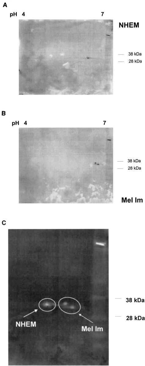
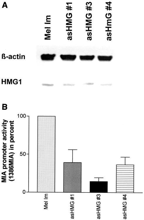
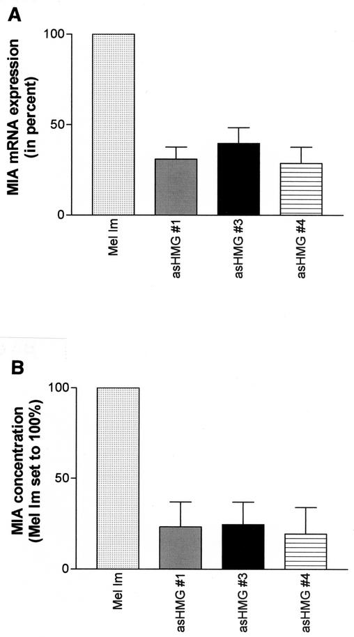
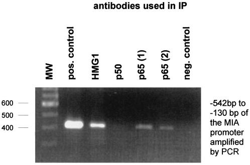
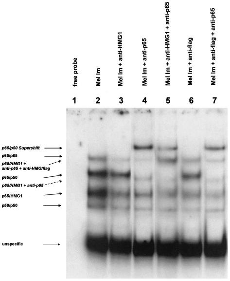
Malignant transformation of melanocytes to melanoma cells closely parallels activation of melanoma inhibitory activity (MIA) expression. We have previously shown that upregulation of MIA occurs on a transcriptional level and involves the highly conserved region (HCR) promoter element. We further observed that the HCR element interacts with the melanoma-associated transcription factor (MATF) and thereby confers strong promoter activation. In this study we identify the peptide sequence of MATF and show that it is identical with the transcription factor HMG1. HMG1 was upregulated in malignant melanoma cells and further activated by hypophosphorylation. Stable antisense-HMG1 expression in melanoma cells led to the reduction of MIA promoter activity and protein expression, indicating that HMG1 is a potent regulator of MIA expression. Interestingly, chromatin immunoprecipitation and electrophoretic mobility shift experiments indicated that HMG1 and the NF-kappa B family member p65 both interact and bind to the HCR promoter element. In summary, our study proves HMG1 and p65 to be important factors in MIA regulation and melanoma progression.


Figures

FIG. 1.
FIG. 1.
Peptide sequences of purified MATF. MATF was purified (8) and cleaved by protease LysC digestion. Peptides were purified by reverse-phase HPLC and sequenced, and two peptides were clearly identified (underlined) as HMG1 fragments (GenBank accession no. X12597).
FIG. 2.
FIG. 2.
HMG1 protein expression in malignant melanoma and NHEM. (A) Western blotting revealed strong upregulation of HMG1 expression in nine melanoma cell lines (Mel Im, Mel Ju, Mel Juso, Mel Ho, Mel Wei, Mel Ei, SK-Mel-3, SK-Mel-28, and HTZ19d) compared to that in NHEM. Seven micrograms of protein lysate per lane was applied. Coincubation with a β-actin antibody (Sigma) was used to ensure equal loadings. (B) Analysis of normal skin by immunohistochemistry revealed very weak HMG1 signals in epidermal melanocytes in the basal layer of the skin (panel II, arrows). Tyrosinase staining in serial sections was used to identify the melanocytes (panel I, arrows). All keratinocytes were found to be highly positive for HMG1. In melanoma cells, significant HMG1 expression was found (panels III and IV).
FIG. 2.
FIG. 2.
HMG1 protein expression in malignant melanoma and NHEM. (A) Western blotting revealed strong upregulation of HMG1 expression in nine melanoma cell lines (Mel Im, Mel Ju, Mel Juso, Mel Ho, Mel Wei, Mel Ei, SK-Mel-3, SK-Mel-28, and HTZ19d) compared to that in NHEM. Seven micrograms of protein lysate per lane was applied. Coincubation with a β-actin antibody (Sigma) was used to ensure equal loadings. (B) Analysis of normal skin by immunohistochemistry revealed very weak HMG1 signals in epidermal melanocytes in the basal layer of the skin (panel II, arrows). Tyrosinase staining in serial sections was used to identify the melanocytes (panel I, arrows). All keratinocytes were found to be highly positive for HMG1. In melanoma cells, significant HMG1 expression was found (panels III and IV).
FIG. 3.
FIG. 3.
Detection of posttranslational modification of HMG1 in malignant melanoma and primary skin melanocytes. By two-dimensional gel electrophoresis and subsequent Western blotting, different electrophoretic mobilities, most likely resulting from a reduction of phosphorylation of HMG1, were detected in melanoma cells (B) and NHEM (A). (C) Overlay to compare the differences in electrophoretic mobilities. As NHEM expressed significantly lower levels of HMG1, a fourfold excess of protein was loaded.
FIG. 4.
FIG. 4.
(A) HMG1 protein levels in stably antisense-HMG1 (asHMG #1, 3, and 4)-transfected cell clones compared to those in the parental cell line Mel Im. Expression of HMG1 protein in the parental cell line Mel Im and in stably antisense-HMG1-transfected cell clones was detected by Western blotting with an HMG1 antibody. HMG1 levels in melanoma cells were reduced by stable transfection of antisense-HMG1 expression constructs. Equal loadings were verified by coincubation of the blotting membrane with a β-actin antibody. (B) MIA promoter activity in antisense-HMG1-transfected cell clones. Analysis by luciferase reporter assays of the antisense-HMG1-transfected cell clones revealed a reduced activity of the 1,386-bp fragment of the MIA promoter (1386MIA).
FIG. 5.
FIG. 5.
MIA mRNA and protein expression in antisense-HMG1 (asHMG #1)-transfected cell clones. HMG1 levels in melanoma cells were reduced by stable transfection of antisense-HMG1 expression constructs. Analysis of the cell clones revealed reduced MIA mRNA and protein expression as measured in tissue culture supernatant by RT-PCR (A) and ELISA (B).
FIG. 6.
FIG. 6.
Interaction of HMG1 and p65 of the NF-κB family with the MIA promoter as assayed by chromatin immunoprecipitation (IP). After bound transcription factors were fixed to the chromatin (see Materials and Methods), DNA was sheared by sonication and protein-DNA complexes were used in different immunoprecipitations (with anti-HMG1, anti-p50, and two different anti-p65 antibodies). Subsequently, the DNA was separated from the precipitated protein complexes and used in a specific PCR amplifying the HCR of the MIA promoter (bp −542 to −130). Chromatin immunoprecipitation proved binding of HMG1 and p65 to the region in the MIA promoter in vivo, whereas no binding of p50 was observed. Molecular weight (MW) markers are shown at the left. pos., positive; neg. negative.
FIG. 7.
FIG. 7.
Analysis of the direct interaction of HMG1 with NF-κB family members by gel mobility shift assays. Nuclear extracts of the melanoma cell line Mel Im (lane 2), transiently transfected with flag-sense-HMG1 expression plasmids, were incubated with the oligomeric HCR-binding site of the MIA promoter. Lanes 3 and 6 show a decrease in the band intensity of the band shift labeled p65/HMG1 after incubation of the nuclear extracts of Mel Im with anti-HMG1 (lane 3) or anti-flag (lane 6). Band shifting of the bands labeled p65/p65, p65/p50, and p65/HMG1 was observed after incubation of the extracts with anti-p65 (lane 4). In lanes 5 and 7, nuclear extracts were coincubated with two different antibodies: anti-HMG1 and anti-p65 (lane 5) or anti-flag and anti-p65 (lane 7). This resulted in the additional supershifted band of the lane labeled p65/HMG1 + anti-p65 + anti-HMG/flag.
FIG. 8.
FIG. 8.
Coimmunoprecipitation assays. Coimmunoprecipitation (IP) was utilized to show direct interaction of HMG1 and p65. For coimmunoprecipitation of HMG1 with p65, two different anti-p65 antibodies were used (see Materials and Methods). p50 coimmunoprecipitation is just feasible with p65 from Santa Cruz, as p65 from Rockland binds to the p50-binding site. Western blots were stained with anti-HMG1 (panel I) and anti-p50 (panel II) antibodies, respectively. The data show that HMG1 is able to bind to p65 of the NF-κB family on the MIA promoter.

References

    1. Baxevanis, A. D., and D. Landsman. 1995. The HMG-1 box protein family: classification and functional relationships. Nucleic Acids Res. 23:1604-1613. - PMC - PubMed
    1. Blesch, A., A. K. Bosserhoff, R. Apfel, C. Behl, B. Hessdoerfer, A. Schmitt, P. Jachimczak, F. Lottspeich, R. Buettner, and U. Bogdahn. 1994. Cloning of a novel malignant melanoma-derived growth-regulatory protein, MIA. Cancer Res. 54:5695-5701. - PubMed
    1. Boonyaratanakornkit, V., V. Melvin, P. Prendergast, M. Altmann, L. Ronfani, M. E. Bianchi, L. Taraseviciene, S. K. Nordeen, E. A. Allegretto, and D. P. Edwards. 1998. High-mobility group chromatin proteins 1 and 2 functionally interact with steroid hormone receptors to enhance their DNA binding in vitro and transcriptional activity in mammalian cells. Mol. Cell. Biol. 18:4471-4487. - PMC - PubMed
    1. Bosserhoff, A. K., A. Hauschild, R. Hein, D. Schadendorf, E. Stockflesh, T. Bogenrieder, M. Landthaler, R. Buettner, and W. Stolz. 2000. Elevated MIA serum levels are of relevance for management of metastasized malignant melanomas: results of a German multicenter study. J. Investig. Dermatol. 114:395-396. - PubMed
    1. Bosserhoff, A. K., R. Hein, U. Bogdahn, and R. Buettner. 1996. Structure and promoter analysis of the gene encoding the human melanoma-inhibiting protein MIA. J. Biol. Chem. 271:490-495. - PubMed

Publication types

MeSH terms