

Cardiovascular and endocrine responses to acute hypoxaemia during and following dexamethasone infusion in the ovine fetus
- PMID: 12665612
- PMCID: PMC2342926
- DOI: 10.1113/jphysiol.2002.036418
Cardiovascular and endocrine responses to acute hypoxaemia during and following dexamethasone infusion in the ovine fetus
Abstract
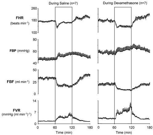
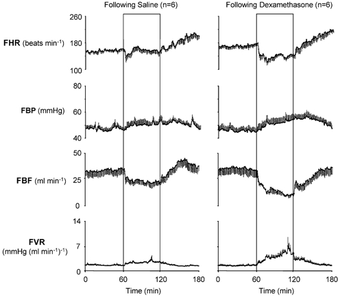
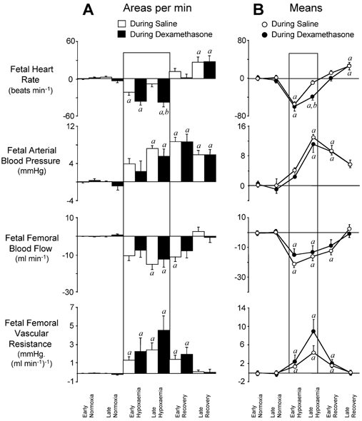
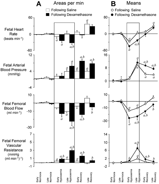
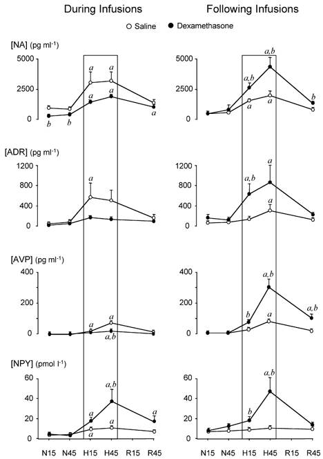
This study investigated the effects of fetal treatment with dexamethasone on ovine fetal cardiovascular defence responses to acute hypoxaemia, occurring either during or 48 h following the period of glucocorticoid exposure. To address the mechanisms underlying these responses, chemoreflex function and plasma concentrations of catecholamines, neuropeptide Y (NPY) and vasopressin were measured. Under general halothane anaesthesia, 26 Welsh Mountain sheep fetuses were surgically prepared for long-term recording at between 117 and 120 days of gestation (dGA; term is approximately 145 days) with vascular catheters and a Transonic flow probe around a femoral artery. Following at least 5 days of recovery, fetuses were randomly assigned to one of two experimental groups. After 48 h of baseline recording, at 125 +/- 1 dGA, half of the fetuses (n = 13) were continuously infused I.V. with dexamethasone for 48 h at a rate of 2.06 +/- 0.13 microg kg-1 h-1. The remaining 13 fetuses were infused with heparinized saline at the same rate (controls). At 127 +/- 1 dGA, 2 days from the onset of infusions, seven fetuses from each group were subjected to 1 h of acute hypoxaemia. At 129 +/- 1 dGA, 2 days after the end of infusions, six fetuses from each group were subjected to 1 h of acute hypoxaemia. Similar reductions in fetal partial pressure of arterial oxygen occurred in control and dexamethasone-treated fetuses during the acute hypoxaemia protocols. In control fetuses, acute hypoxaemia led to transient bradycardia, femoral vasoconstriction and significant increases in plasma concentrations of catecholamines, vasopressin and NPY. In fetuses subjected to acute hypoxaemia during dexamethasone treatment, the increase in plasma NPY was enhanced, the bradycardic response was prolonged, and the plasma catecholamine and vasopressin responses were diminished. In fetuses subjected to acute hypoxaemia 48 h following dexamethasone treatment, femoral vasoconstriction and plasma catecholamine and vasopressin responses were enhanced, whilst the prolonged bradycardia and augmented plasma NPY responses persisted. These data show that fetal treatment with dexamethasone modifies the pattern and magnitude of fetal cardiovascular responses to acute oxygen deprivation. Modifications to different mechanisms mediating the fetal defence responses to acute hypoxaemia that occur during dexamethasone treatment may reverse, persist or even become enhanced by 48 h following the treatment period.
Figures
Comment in
-
Fetal exposure to corticosteroids: how low can we go?J Physiol. 2003 May 15;549(Pt 1):1. doi: 10.1113/jphysiol.2003.041459. Epub 2003 Mar 28. J Physiol. 2003. PMID: 12665601 Free PMC article. Review. No abstract available.
Similar articles
-
Effects of low dose dexamethasone treatment on basal cardiovascular and endocrine function in fetal sheep during late gestation.J Physiol. 2002 Dec 1;545(2):649-60. doi: 10.1113/jphysiol.2001.015693. J Physiol. 2002. PMID: 12456840 Free PMC article.
-
Fetal cardiovascular, metabolic and endocrine responses to acute hypoxaemia during and following maternal treatment with dexamethasone in sheep.J Physiol. 2005 Sep 1;567(Pt 2):673-88. doi: 10.1113/jphysiol.2005.089805. Epub 2005 Jun 23. J Physiol. 2005. PMID: 15975982 Free PMC article.
-
Enhanced nitric oxide activity offsets peripheral vasoconstriction during acute hypoxaemia via chemoreflex and adrenomedullary actions in the sheep fetus.J Physiol. 2003 Feb 15;547(Pt 1):283-91. doi: 10.1113/jphysiol.2002.032615. Epub 2003 Jan 10. J Physiol. 2003. PMID: 12562956 Free PMC article.
-
The fetal llama versus the fetal sheep: different strategies to withstand hypoxia.High Alt Med Biol. 2003 Summer;4(2):193-202. doi: 10.1089/152702903322022794. High Alt Med Biol. 2003. PMID: 12855051 Review.
-
Human cardiovascular adjustments to acute hypoxaemia.Clin Physiol. 1987 Oct;7(5):349-76. doi: 10.1111/j.1475-097x.1987.tb00179.x. Clin Physiol. 1987. PMID: 3311579 Review.
Cited by
-
Perinatal glucocorticoid sensitivity in the preterm newborn: molecular mechanisms, endogenous determinants, and clinical implications.Front Endocrinol (Lausanne). 2025 Jul 16;16:1587891. doi: 10.3389/fendo.2025.1587891. eCollection 2025. Front Endocrinol (Lausanne). 2025. PMID: 40741174 Free PMC article. Review.
-
Breath of Life: Heart Disease Link to Developmental Hypoxia.Circulation. 2021 Oct 26;144(17):1429-1443. doi: 10.1161/CIRCULATIONAHA.121.054689. Epub 2021 Oct 25. Circulation. 2021. PMID: 34694887 Free PMC article.
-
Sodium dichloroacetate stimulates cardiac mitochondrial metabolism and improves cardiac conduction in the ovine fetus during labor.Am J Physiol Regul Integr Comp Physiol. 2022 Jan 1;322(1):R83-R98. doi: 10.1152/ajpregu.00185.2021. Epub 2021 Dec 1. Am J Physiol Regul Integr Comp Physiol. 2022. PMID: 34851727 Free PMC article.
-
Fetal cardiovascular response to acute hypoxia during maternal anesthesia.Physiol Rep. 2020 Feb;8(3):e14365. doi: 10.14814/phy2.14365. Physiol Rep. 2020. PMID: 32026576 Free PMC article.
-
Altered Cardiovascular Defense to Hypotensive Stress in the Chronically Hypoxic Fetus.Hypertension. 2020 Oct;76(4):1195-1207. doi: 10.1161/HYPERTENSIONAHA.120.15384. Epub 2020 Aug 31. Hypertension. 2020. PMID: 32862711 Free PMC article.
References
-
- Adams MB, Ross JT, Butler TG, McMillen IC. Glucocorticoids decrease phenylethanolamine N-methyltransferase mRNA expression in the immature foetal sheep adrenal. J Neuroendocrinol. 1999;11:569–575. - PubMed
-
- Akagi K, Berdusco ET, Challis JR. Cortisol inhibits ACTH but not the AVP response to hypoxaemia in fetal lambs at days 123–128 of gestation. J Dev Physiol. 1990;14:319–324. - PubMed
-
- Alexander DP, Bashore RA, Britton HG, Forsling ML. Maternal and fetal arginine vasopressin in the chronically catheterised sheep. Biol Neonate. 1974;25:242–248. - PubMed
-
- Allen JM, Adrian TE, Polak JM, Bloom SR. Neuropeptide Y (NPY) in the adrenal gland. J Auton Nerv Syst. 1983;9:559–563. - PubMed
-
- Altura BM, Altura BT. Peripheral vascular actions of glucocorticoids and their relationship to protection in circulatory shock. J Pharmacol Exp Ther. 1974;190:300–315. - PubMed
Publication types
MeSH terms
Substances
LinkOut - more resources
Full Text Sources
Medical
Miscellaneous