. 2003 Apr 15;100(8):4754-9.
doi: 10.1073/pnas.0730743100. Epub 2003 Mar 28.

Smooth muscle cells in human coronary atherosclerosis can originate from cells administered at marrow transplantation

Affiliations

Smooth muscle cells in human coronary atherosclerosis can originate from cells administered at marrow transplantation

Noel M Caplice et al. Proc Natl Acad Sci U S A. .

Abstract

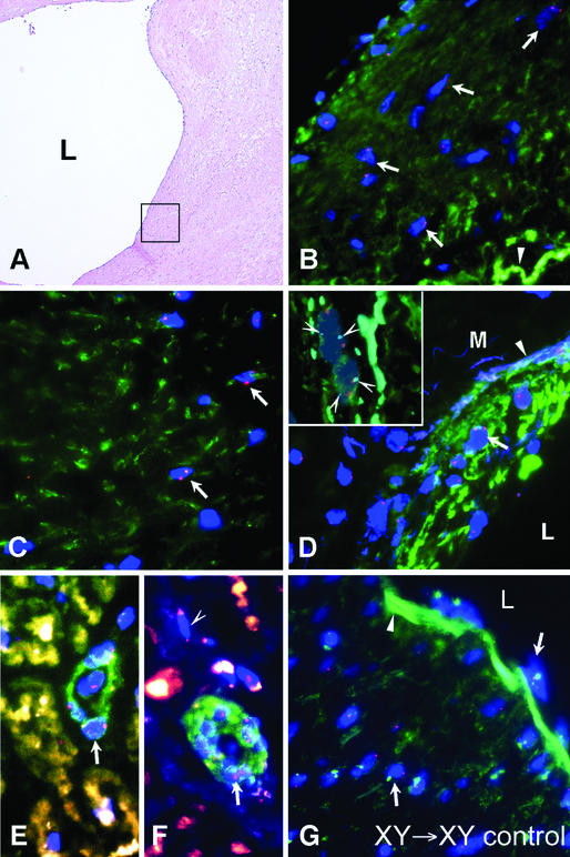
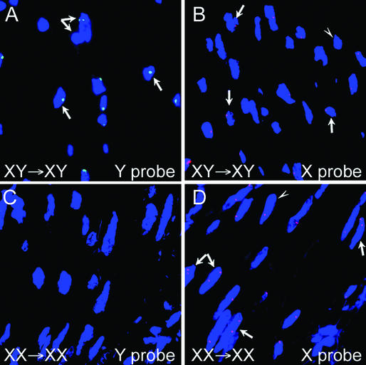
Atherosclerosis is the major cause of adult mortality in the developed world, and a significant contributor to atherosclerotic plaque progression involves smooth muscle cell recruitment to the intima of the vessel wall. Controversy currently exists on the exact origin of these recruited cells. Here we use sex-mismatched bone marrow transplant subjects to show that smooth muscle cells throughout the atherosclerotic vessel wall can derive from donor bone marrow. We demonstrate extensive recruitment of these cells in diseased compared with undiseased segments and exclude cell-cell fusion events as a cause for this enrichment. These data have broad implications for our understanding of the cellular components of human atherosclerotic plaque and provide a potentially novel target for future diagnostic and therapeutic strategies.


Figures

Figure 1
Figure 1
Intimal atherosclerotic plaque, media, and adventitial microvessels from female recipients (subjects 1–4, Table 1) who received a male BMT (XY → XX; AH). (A) Hematoxylin and eosin staining showing a grade III atherosclerotic plaque from subject 1. L, lumen; filled arrowhead, internal elastic lamina. The two boxes indicate the subendothelial intima and the deep intima that are subsequently analyzed with dual α smooth muscle actin staining and FISH for Y chromosome in B and C. (B) Combined immunostaining for α smooth muscle actin and FISH for Y chromosome showing clusters of male cells (blue DAPI-stained nuclei with green dot) surrounded by positive α smooth muscle actin staining (red Cy-3 stain surrounding male nuclei, white arrows) in the subendothelial intima of plaque from subject 1. A row of white arrows point to a cluster of male actin-positive cells. Some actin staining cells were negative for the Y chromosome (open arrowhead). (C) Male smooth muscle cells deeper within the intima of plaque from subject 1 colabeling for α smooth muscle actin and the Y chromosome (white arrows). (D) Male smooth muscle cells in the media of coronary artery from subject 2 showing combined labeling for α smooth muscle actin and the Y chromosome (white arrows). The open arrowhead indicates the elastic lamellae of the media (M) and the filled arrowhead indicates the internal elastic lamina. (Inset) Chromosomal multiploidy analysis of male donor smooth muscle cells in female coronary artery showing diploid nature and lack of cell fusion. Shown are two separate nuclei (DAPI) each showing single X chromosome (large red dot) and Y chromosome (green dot) and also two chromosome 18 markers per nucleus (light blue dots, arrowheads), indicating each nucleus is diploid. The background red staining indicates α smooth muscle actin staining with Cy-3 fluorescence. (E and F) Male smooth muscle cells within the subendothelial and deep intima of a coronary artery plaque from subject 4 showing combined labeling for smooth muscle myosin heavy chain (E, red staining) and smooth muscle calponin (F, red staining) and the Y chromosome (white arrows). (G and H) Male smooth muscle cells in microvessels of the adventitia of an atherosclerotic coronary artery from subject 3 showing combined labeling for α smooth muscle actin and the Y chromosome (white arrows). The open arrowhead indicates an actin staining cell that was negative for the Y chromosome. (I) Combined FISH for X and Y chromsome and α smooth muscle actin costaining showing absence of male cells in a control diseased artery of a same-gender female BMT subject. In BI the nuclei were counterstained with DAPI (blue).
Figure 2
Figure 2
Infrequent donor cells detected in the artery wall of undiseased vessel segments from male and female subjects with gender-mismatched BMT. (A) Hematoxylin and eosin staining of undiseased vessel segment from female recipient who had gender-mismatched BMT. The box indicates an area of intima and media and adventitia subsequently analyzed in C by using α smooth muscle actin-Cy3 staining and FISH for X and Y chromosomes. (B) Hematoxylin and eosin staining of undiseased branch vessel segment from male subject who had gender-mismatched BMT. The box indicates an area of the intima and media subsequently analyzed in D by using α smooth muscle actin-AlexaFluor staining and FISH for the X and Y chromosomes. (C) Lack of male smooth muscle cells in the wall of an undiseased vessel segment in same female subject as in A. Note multiple XX chromosome positive female nuclei (open arrowheads) throughout the vessel and a solitary Y chromosome positive nucleus (arrow) in the adventitia. (D) Infrequent female donor smooth muscle cells in the intima and media of an undiseased vessel segment in the same male subject as in B. Note single XX positive nucleus (two red dots, arrow) in the media where multiple XY positive (red and green dot) male cells are present. In all four panels, an arrowhead indicates the internal elastic lamina.
Figure 3
Figure 3
Intimal atherosclerotic plaque, media, diffuse intimal thickening and adventitial microvessels from male recipients (subjects 5–8, Table 1) who received a female BMT (XX → XY; AF). (A) Hematoxylin and eosin staining showing a grade III atherosclerotic plaque from subject 5. L indicates the lumen and the box indicates the subendothelial intima subsequently analyzed in B with dual α smooth muscle actin staining and FISH for the X chromosome. (B) Combined immunostaining for α smooth muscle actin and FISH for the XX chromosome showing female cells (blue nuclei with two red dots) surrounded by positive α smooth muscle actin staining (green AlexaFluor staining surrounding female nuclei; white arrows) scattered throughout the subendothelial intima of plaque from subject 5. The arrowhead indicates the internal elastic lamina. (C) Female smooth muscle cells deeper within the intima of plaque from subject 6 colabeling for α smooth muscle actin and the XX chromosome (white arrows). (D) Female smooth muscle cells within the intima of a coronary artery with diffuse intimal thickening from subject 8 showing combined labeling for α smooth muscle actin and the XX chromosome (white arrow). L indicates the lumen and the arrowhead indicates the internal elastic lamina. (Inset) Chromosomal multiploidy analysis of female donor smooth muscle cells in male coronary artery showing diploid nature. Two separate female nuclei (DAPI), each showing XX chromosomes (two red dots per cell), no Y chromosome, and two chromosome 18 markers (light blue dots, arrowheads) per nucleus indicating diploid status. The green staining surrounding both nuclei indicates α smooth muscle actin labeling with AlexaFluor-conjugated antibody. (E and F) Female smooth muscle cells in microvessels of the adventitia of an atherosclerotic coronary artery from subject 7 showing combined labeling for α smooth muscle actin and the XX chromosome (white arrows). The open arrowhead indicates a female cell not costaining for actin. (G) Control male recipient of same-gender BMT showing absence of female (XX) cells in the intima and media of the vessel wall. Arrows indicate male nuclei and a filled arrowhead indicates the internal elastic lamina. In BG the nuclei were counterstained with DAPI (blue).
Figure 4
Figure 4
No chimeric cells are detected in the vessel wall of same-gender BMT subjects. (A) FISH for male cells showing Y chromosome body (green dot in blue nuclei) in coronary atherosclerotic plaque from a male subject who had same-gender BMT. The arrows indicate male cells with the dual arrow showing two overlapping male nuclei. (B) FISH for female cells in coronary atherosclerotic plaque from the same male subject as in A showing single X chromosome (red dot in nuclei) in each of the intimal cells. The arrows indicate the single X chromosome cells and the arrowhead indicates a cell negative for the X chromosome. No XX chromosome was detected in cells of this subject. (C) FISH for male cells in media of diffuse intimal thickening from a female subject who had same-gender BMT showing lack of Y chromosome labeling. (D) FISH for female cells showing XX chromosome labeling (two red dots per nucleus; arrows) in cells in the media of diffuse intimal thickening from same female subject as in C. The presence of single red dots in some nuclei (arrowhead) indicates that not all cells labeled positive for the XX chromosome.

References

    1. Duguid J B, Robertson W B. Lancet. 1957;1:1205–1209. - PubMed
    1. Ross R, Glomset J A. Science. 1973;180:1332–1339. - PubMed
    1. Virchow R. Wien Med Wochenschr. 1856;6:825–829.
    1. von Rokitansky C. A Manual of Pathological Anatomy. Berlin: Berlin Sydenham Society; 1852.
    1. Ross R, Glomset J A. N Engl J Med. 1976;295:369–377. - PubMed

Publication types