

Epstein-Barr virus-infected B cells expanding in germinal centers of infectious mononucleosis patients do not participate in the germinal center reaction
- PMID: 12665622
- PMCID: PMC153624
- DOI: 10.1073/pnas.2627966100
Epstein-Barr virus-infected B cells expanding in germinal centers of infectious mononucleosis patients do not participate in the germinal center reaction
Abstract
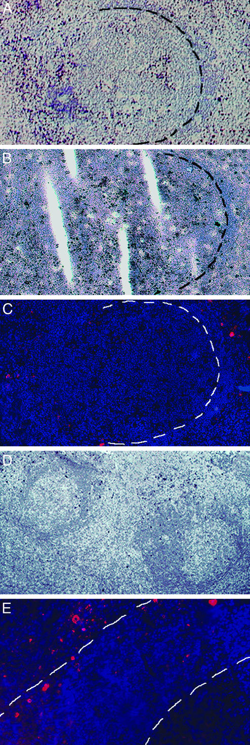
To assess the impact of the germinal center (GC) reaction on viral spread in Epstein-Barr virus (EBV) infection, we isolated EBV(+) GC B cells from the tonsils of two infectious mononucleosis patients, sequenced their rearranged V genes, and determined expression of the EBV latency genes EBV nuclear antigen 2 and latent membrane protein 1. Most EBV(+) GC B cells belonged to clones of cells harboring somatically mutated V gene rearrangements. Ongoing somatic hypermutation, the hallmark of the GC reaction, was seen only in uninfected GC B cell clones, not in EBV(+) B cell clones. Thus, in infectious mononucleosis, GC and/or memory B cells are directly infected by EBV and expand without somatic hypermutation, whereas the GC passage of EBV-infected naive B cells does not contribute detectably to the generation of infected memory B cells, the main reservoir of EBV during persistence. Most, if not all, EBV-infected cells in GCs exhibited an unusual EBV gene expression pattern in that they were positive for EBV nuclear antigen 2 but negative for latent membrane protein 1. Although the three main types of EBV-associated B cell lymphomas (Burkitt's, Hodgkin's, and posttransplant lymphomas) presumably are derived from GC B cells, EBV(+) GC B cells resembling these EBV(+) GC B cell lymphomas in terms of EBV gene expression and somatic hypermutation pattern could not be identified.
Figures
References
-
- Rickinson A B, Kieff E. In: Fields Virology. Knipe D M, Howley P M, editors. Philadelphia: Lippincott; 2001. pp. 2575–2627.
-
- Caldwell R G, Wilson J B, Anderson S J, Longnecker R. Immunity. 1998;9:405–411. - PubMed
-
- Zimber-Strobl U, Strobl L J. Semin Cancer Biol. 2001;11:423–434. - PubMed
-
- Babcock G J, Hochberg D, Thorley-Lawson A D. Immunity. 2000;13:497–506. - PubMed
Publication types
MeSH terms
Substances
Associated data
- Actions
- Actions
- Actions
- Actions
- Actions
- Actions
- Actions
- Actions
- Actions
- Actions
- Actions
- Actions
- Actions
- Actions
- Actions
- Actions
- Actions
- Actions
- Actions
- Actions
- Actions
- Actions
- Actions
- Actions
- Actions
- Actions
- Actions
- Actions
- Actions
- Actions
- Actions
- Actions
- Actions
- Actions
- Actions
- Actions
- Actions
- Actions
- Actions
- Actions
- Actions
- Actions
- Actions
- Actions
- Actions
- Actions
- Actions
- Actions
- Actions
- Actions
- Actions
- Actions
- Actions
- Actions
- Actions
- Actions
- Actions
- Actions
- Actions
- Actions
- Actions
- Actions
- Actions
- Actions
- Actions
- Actions
- Actions
- Actions
- Actions
- Actions
- Actions
- Actions
- Actions
- Actions
- Actions
- Actions
- Actions
- Actions
- Actions
- Actions
- Actions
- Actions
- Actions
- Actions
- Actions
- Actions
- Actions
- Actions
- Actions
- Actions
- Actions
- Actions
- Actions
- Actions
- Actions
- Actions
- Actions
- Actions
- Actions
- Actions
- Actions
- Actions
- Actions
- Actions
- Actions
- Actions
- Actions
- Actions
- Actions
- Actions
- Actions
- Actions
- Actions
- Actions
- Actions
- Actions
- Actions
- Actions
- Actions
- Actions
- Actions
- Actions
- Actions
- Actions
- Actions
- Actions
- Actions
- Actions
- Actions
- Actions
- Actions
- Actions
- Actions
- Actions
- Actions
- Actions
- Actions
- Actions
- Actions
- Actions
- Actions
- Actions
- Actions
- Actions
- Actions
- Actions
- Actions
- Actions
- Actions
- Actions
- Actions
- Actions
- Actions
- Actions
- Actions
- Actions
- Actions
- Actions
- Actions
- Actions
- Actions
- Actions
- Actions
- Actions
- Actions
- Actions
- Actions
- Actions
- Actions
- Actions
- Actions
- Actions
- Actions
- Actions
- Actions
- Actions
- Actions
- Actions
- Actions
- Actions
- Actions
- Actions
- Actions
- Actions
- Actions
- Actions
- Actions
- Actions
- Actions
- Actions
- Actions
- Actions
- Actions
- Actions
- Actions
- Actions
- Actions
- Actions
- Actions
- Actions
- Actions
- Actions
- Actions
- Actions
- Actions
- Actions
- Actions
- Actions
- Actions
- Actions
- Actions
- Actions
- Actions
- Actions
- Actions
- Actions
- Actions
- Actions
- Actions
- Actions
- Actions
- Actions
- Actions
- Actions
- Actions
- Actions
- Actions
- Actions
- Actions
- Actions
- Actions
- Actions
- Actions
- Actions
- Actions
- Actions
- Actions
- Actions
- Actions
- Actions
- Actions
- Actions
- Actions
- Actions
- Actions
- Actions
- Actions
- Actions
- Actions
- Actions
- Actions
- Actions
- Actions
- Actions
- Actions
- Actions
- Actions
- Actions
- Actions
LinkOut - more resources
Full Text Sources
Medical
Molecular Biology Databases
Research Materials
Miscellaneous
