. 2003 Apr;137(1-2):42-50.
doi: 10.1016/s0165-5728(03)00041-9.

Bystander CD4 T cells do not mediate demyelination in mice infected with a neurotropic coronavirus

Affiliations

Bystander CD4 T cells do not mediate demyelination in mice infected with a neurotropic coronavirus

Jodie S Haring et al. J Neuroimmunol. 2003 Apr.

Abstract

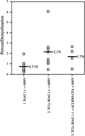
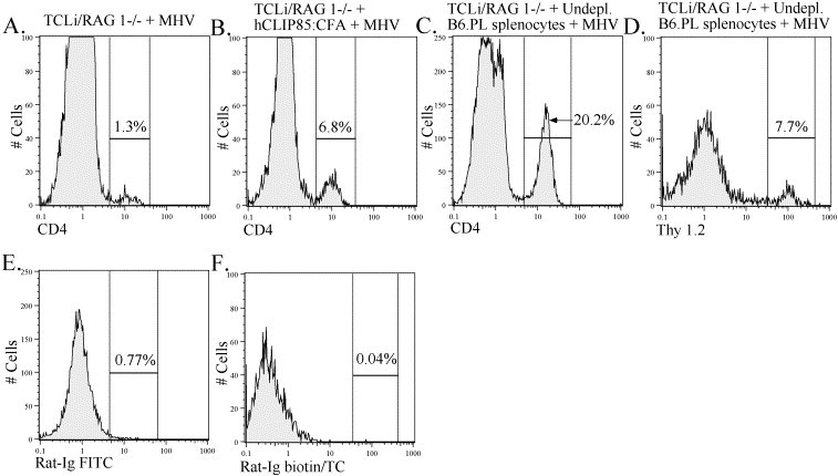
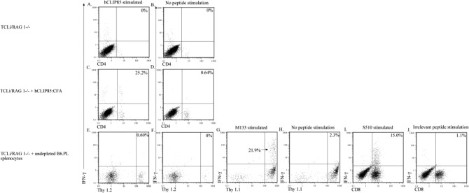
Demyelination following infection of mice with the neurotropic coronavirus mouse hepatitis virus strain JHM (MHV) is immune-mediated. It has been demonstrated that MHV-specific CD4 and CD8 T cells are capable of causing demyelination independent of the other T cell subset. Recent work has also demonstrated that activated bystander CD8 T cells mediate significant demyelination. The ability of bystander CD4 T cells to mediate demyelination was investigated using CD4 T cell transgenic mice. The results indicated that bystander CD4 T cells were unable to cause demyelination in MHV-infected mice, despite being recruited into the central nervous system (CNS) and irrespective of activation status. These results suggest that CD4 T cells must recognize antigen in the CNS in order to cause demyelination.


Figures

Fig. 1
Fig. 1
Bystander CD4 T cells were unable to cause demyelination. Little demyelination was detected in MHV-infected RAG1−/− mice (column 1), in MHV-infected TCLi mice (column 2), or in hCLIP85:CFA-treated, MHV-infected TCLi mice (column 3). Black horizontal lines in each column represent the mean percent demyelination for each group. Demyelination±S.D. was 0.7±0.2%, 2.1±0.5% and 1.7±0.5% for Groups 1, 2 and 3, respectively. Demyelination values in columns 2 and 3 are not statistically different from control values in column 1.
Fig. 2
Fig. 2
Bystander CD4 T cell recruitment into the CNS was increased in the presence of other T cells and after in vivo activation with cognate peptide. Lymphocytes were isolated from the brains of TCLi mice that were infected with MHV (A), infected TCLi mice that were previously treated with hCLIP85:CFA (B, E) and infected TCLi mice that received 5×10⁶ undepleted splenocytes from an MHV-immunized B6.PL mouse (C, D, F). Lymphocytes were stained for CD4 and Thy 1.2 (transgenic CD4 T cell marker) as described in Section 2. Lymphocytes from individual animals were analyzed. Histograms shown are representative of results obtained from five TCLi-infected mice, four hCLIP85:CFA-treated, infected TCLi mice and eight infected TCLi mice receiving adoptively transferred splenocytes. Percentages represent the percent of lymphocytes that fall within the indicated gate. Panels D and F were gated on CD4+ cells.
Fig. 3
Fig. 3
Bystander activation of TCLi CD4 T cells as measured by intracellular cytokine staining was minimal. Lymphocytes were isolated from the brains of mice in each of the experimental groups listed at the left of each row. Cells were stained for intracellular IFN-γ as described in Section 2. The peptide used to stimulate the cells in each panel is indicated at the top of each column. Panels A, C and E show staining of TCLi transgenic T cells after in vitro stimulation with hCLIP85. Panels B, D and F show staining of TCLi cells with no peptide stimulation and serve as controls for panels A, C and E. Panel G shows staining of transferred B6.PL CD4 T cells after stimulation with MHV-specific CD4 T cell peptide M133. Panel H shows staining of B6.PL CD4 T cells with no peptide stimulation and serves a control for G. Panels E–H are gated on CD4+ T cells. Panel I shows staining of B6.PL CD8 T cells after stimulation with MHV-specific CD8 T cell peptide S510. Panel J shows staining of B6.PL CD8 T cells after stimulation with an irrelevant peptide (Ova257) and serves as a control for panel I. Percentages are the percent of CD4 or CD8 T cells that fall in the indicated quadrant.

References

    1. Bauer J., Bradl M., Hickey W., Forss-peter S., Breitscopf H., Linington C., Wekerle H., Lassmann H. T-cell apoptosis in inflammatory brain lesions: destruction of T cells does not depend on antigen recognition. Am. J. Pathol. 1998;153:715–724. - PMC - PubMed
    1. Brabb T., von Dassow P., Ordonez N., Schnabel B., Duke B., Goverman J. In situ tolerance within the central nervous system as a mechanism for preventing autoimmunity. J. Exp. Med. 2000;192:871–880. - PMC - PubMed
    1. Brehm M.A., Pinto A.K., Daniels K.A., Schneck J.P., Welsh R.M., Selin L.K. T cell immunodominance and maintenance of memory regulated by unexpectedly cross-reactive pathogens. Nat. Immunol. 2002;3:627–634. - PubMed
    1. Deshpande S., Zheng M., Lee S., Banerjee K., Gangappa S., Kumaraguru U., Rouse B. Bystander activation involving T lymphocytes in herpetic stromal keratitis. J. Immunol. 2001;167:2902–2910. - PubMed
    1. Ehl S., Hombach J., Aichele P., Hengartner H., Zinkernagel R.M. Bystander activation of cytotoxic T Cells: studies on the mechanism and evaluation of in vivo significance in a transgenic mouse model. J. Exp. Med. 1997;185:1241–1251. - PMC - PubMed