Comparative Study
. 2003 Apr;84(4):2700-8.
doi: 10.1016/S0006-3495(03)75075-9.

Extra spike formation in sensory neurons and the disruption of afferent spike patterning

Affiliations
Comparative Study

Extra spike formation in sensory neurons and the disruption of afferent spike patterning

Ron Amir et al. Biophys J. 2003 Apr.

Abstract

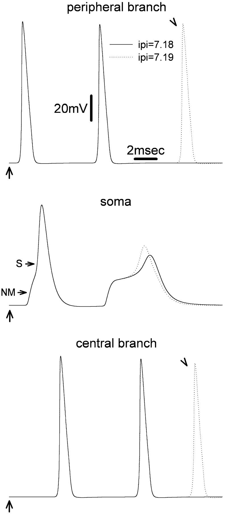
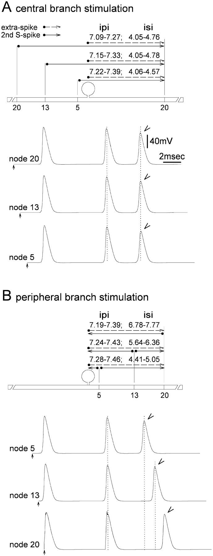
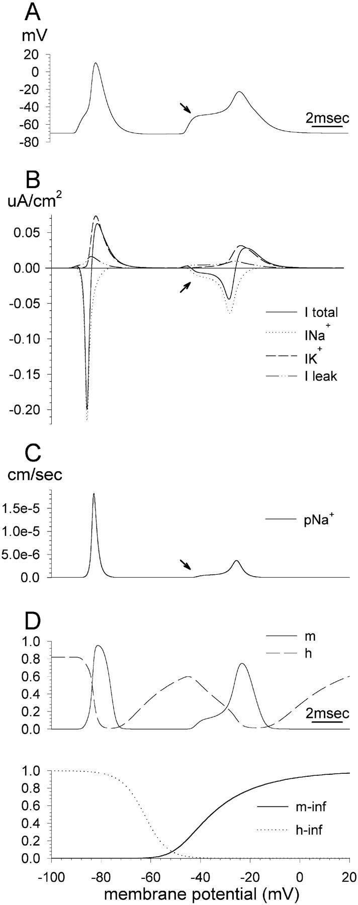
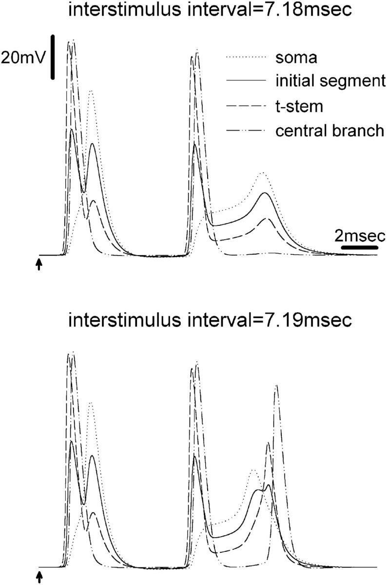
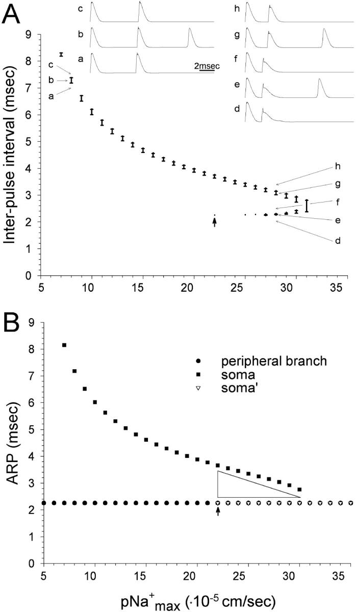
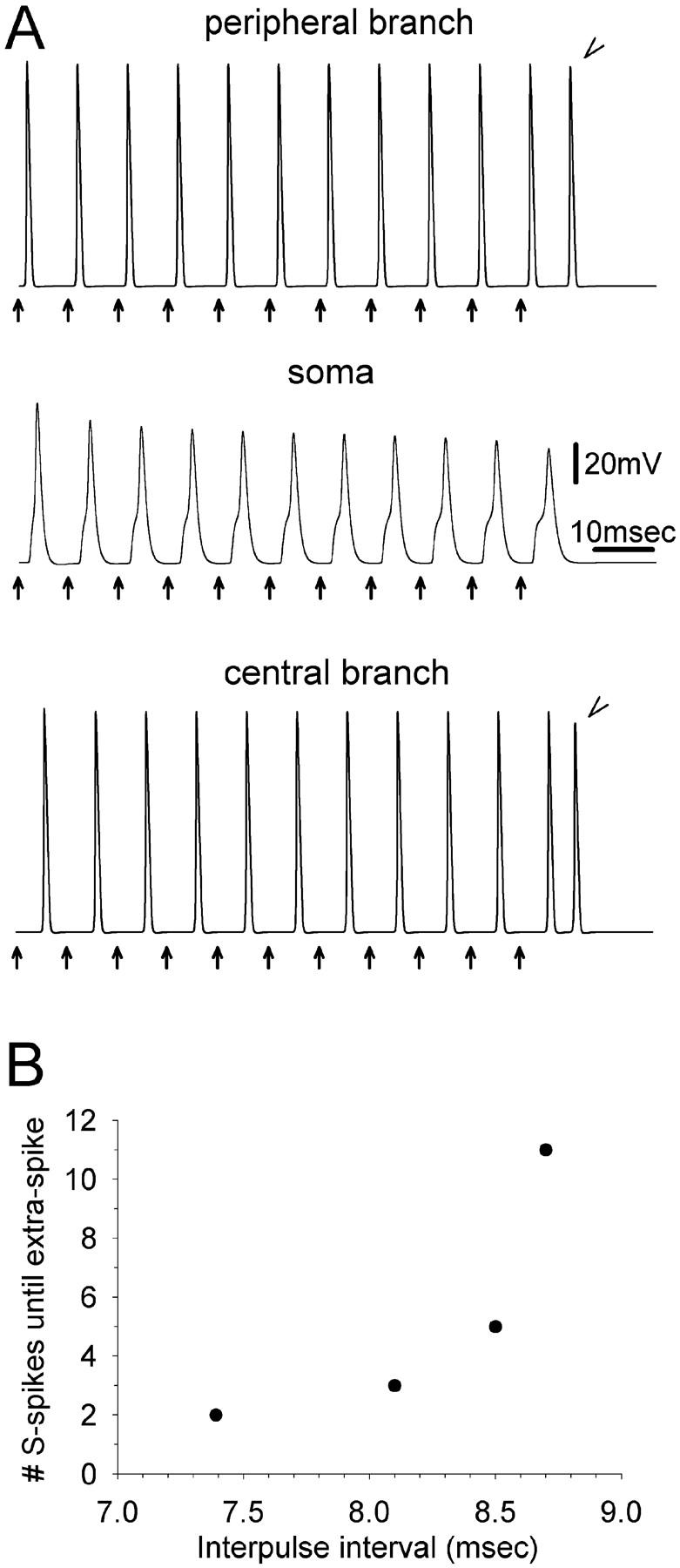
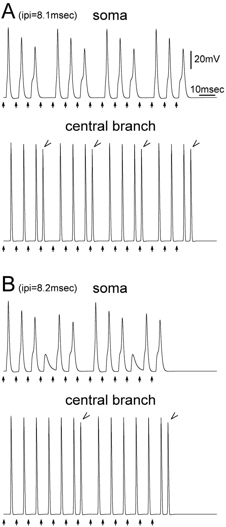
The peculiar pseudounipolar geometry of primary sensory neurons can lead to ectopic generation of "extra spikes" in the region of the dorsal root ganglion potentially disrupting the fidelity of afferent signaling. We have used an explicit model of myelinated vertebrate sensory neurons to investigate the location and mechanism of extra spike formation, and its consequences for distortion of afferent impulse patterning. Extra spikes originate in the initial segment axon under conditions in which the soma spike becomes delayed and broadened. The broadened soma spike then re-excites membrane it has just passed over, initiating an extra spike which propagates outwards into the main conducting axon. Extra spike formation depends on cell geometry, electrical excitability, and the recent history of impulse activity. Extra spikes add to the impulse barrage traveling toward the spinal cord, but they also travel antidromically in the peripheral nerve colliding with and occluding normal orthodromic spikes. As a result there is no net increase in afferent spike number. However, extra spikes render firing more staccato by increasing the number of short and long interspike intervals in the train at the expense of intermediate intervals. There may also be more complex changes in the pattern of afferent spike trains, and hence in afferent signaling.


Figures

FIGURE 1
FIGURE 1
Extra spikes are observed in the peripheral and the central axon branches at particular values of interpulse interval (IPI). The first of two stimulus pulses (arrows) was delivered at the 20th peripheral node. The second was given 7.18 ms (solid line) or 7.19 ms later (dotted line). Recordings were made from: the most distal node (34th) of the peripheral branch (upper traces), the cell soma (middle traces), and the most central (34th node) of the central branch (bottom traces). Using IPI = 7.18 ms, two spikes were evoked. Increasing IPI to 7.19 ms yielded an extra (third) spike in both the peripheral and the central branch (arrowheads), but not in the cell soma.
FIGURE 2
FIGURE 2
Evidence that extra spikes are generated in the region of the DRG. (A) Stimulating the central branch axon and recording from the peripheral, the delay between the second S-spike and the extra spikes (arrowhead) is independent of the exact site of stimulation (20th, 13th, or fifth central node). (B) Stimulating and recording on the peripheral branch axon, the delay between the second S-spike and extra spikes (arrowhead) varies with the exact site of stimulation (20th, 13th, or fifth peripheral node). This behavior is a consequence of the spike propagation path, given the initiation of extra spikes in the DRG (horizontal solid and dashed lines above sketches of the neuron). Similar observations were made in live DRG neurons by Tagini and Camino (1973).
FIGURE 3
FIGURE 3
Biophysical events underlying the onset of the first and second soma spikes. The first stimulus of the pulse pair was applied at the beginning of the trace and the second 7.19 ms later. (A) Membrane potential recorded in the soma. (B) Transmembrane currents recorded in the soma. (C) Na+ ion permeability in the soma. (D) Values of the gating parameters m and h. Arrows indicate the onset of the second S-spike.
FIGURE 4
FIGURE 4
Membrane potential at various locations during the onset of extra spikes. The first stimulus pulse is indicated by an arrow. The second was given 7.18 (top) or 7.19 ms (bottom) later. Recordings were made from four points: the soma, mid-initial segment, the third node on the t-stem axon, and the fifth node from the t-junction along the central axon branch. With IPI = 7.18 ms, the second S-spike decayed as it propagated along the stem axon; no extra spike was recorded in the central or peripheral axon branch. Increasing IPI to 7.19 ms, a propagated extra spike was present in the central or peripheral axons.
FIGURE 5
FIGURE 5
Extra spikes are generated in the t-stem axon over a broader range of IPI values than support propagation into the main conducting axons. The two top horizontal lines indicate the range of IPI values for which a second NM- and S-spike were present in the cell soma. The remaining horizontal lines show the range of IPI values for which a criterion extra spike was present at the recording locations marked on the left. Insets show transmembrane currents at the first node of the stem axon using IPI = 7.18 ms (upper) and IPI = 7.19 ms (lower). The net current (I total) first became inward for IPI = 7.19 ms, the minimum IPI value for evoking propagated extra spikes.
FIGURE 6
FIGURE 6
With increasing electrical excitability (pNamax+) extra spikes are generated using progressively shorter IPI values. Two successive stimuli were applied to the peripheral axon branch at the 20th node. (A) The range of IPI values that support extra spikes is indicated by vertical bars. For pNamax+ 22–31 cm/s extra spikes were evoked in two separate IPI ranges. Traces ac (pNamax+ = 8 × 10⁻⁵ cm/s) and dh (pNamax+ = 28 × 10−5 cm/s) illustrate zones with a single, and dual extra spike range. For pNamax+ < 7 × 10⁻⁵ cm/s and >31 × 10−5 cm/s extra spikes could not be evoked. (B) Recording from the soma and the peripheral axon branch, ARP for axonal propagation was unchanged over the entire range of pNamax+ values. ARP values for the soma declined with increasing pNamax+. For pNamax+≥22 × 10−5 cm/s ARP for the soma took the same value as the axon. See explanation for the difference between soma and soma′ in Results. The arrow at ARP = 22 × 10−5 cm/s indicates a discontinuity in the soma's ARP function. The physiological value of pNamax+ is ∼8 × 10⁻⁵ cm/s.
FIGURE 7
FIGURE 7
Extra spiking during tetanic stimulation. (A) Eleven stimulus pulses were delivered at the 20th peripheral node (arrows, IPI = 8.7 ms). Recordings were made from the most distal node of the peripheral axon branch (top), the soma (middle), and the last node of the central axon branch (bottom). During the tetanus, onset of the S-spike was progressively delayed. The eleventh stimulus evoked an extra spike, recorded in both the central and the peripheral branch (arrowheads). (B) Using stimulus trains, extra spikes are supported at progressively greater values of IPI.
FIGURE 8
FIGURE 8
Extra spiking disrupts rhythmic firing patterns. Fifteen stimuli (arrows) were given at IPI = 8.1 ms (A) or 8.2 ms (B). Recordings were made from the soma and from the last node of the central axon branch. In A, an extra spike was inserted every fourth spike in the train (arrowheads). In B extra spikes occurred every seventh spike (arrowheads).

References

    1. Amir, R., and M. Devor. 1996. Chemically-mediated cross-excitation in rat dorsal root ganglia. J. Neurosci. 16:4733–4741. - PMC - PubMed
    1. Amir, R., and M. Devor. 2003. Electrical excitability of the soma of sensory neurons is required for spike invasion of the soma, but not for through-conduction. Biophys. J. 84:2181–2191. - PMC - PubMed
    1. Amir, R., M. Michaelis, and M. Devor. 1999. Membrane potential oscillations in dorsal root ganglion neurons: role in normal electrogenesis and in neuropathic pain. J. Neurosci. 19:8589–8596. - PMC - PubMed
    1. Amir, R., M. Michaelis, and M. Devor. 2002. Burst discharge in primary sensory neurons: triggered by subthreshold oscillations, maintained by depolarizing afterpotential. J. Neurosci. 22:1187–1198. - PMC - PubMed
    1. Baccus, S. A. 1998. Synaptic facilitation by reflected action potentials: enhancement of transmission when nerve impulses reverse direction at axon branch points. Proc. Natl. Acad. Sci. USA. 95:8345–8350. - PMC - PubMed

Publication types

MeSH terms

LinkOut - more resources