Comparative Study
. 2003 Mar 31;160(7):1009-15.
doi: 10.1083/jcb.200211091.

uPARAP/Endo180 is essential for cellular uptake of collagen and promotes fibroblast collagen adhesion

Affiliations
Comparative Study

uPARAP/Endo180 is essential for cellular uptake of collagen and promotes fibroblast collagen adhesion

Lars H Engelholm et al. J Cell Biol. .

Abstract

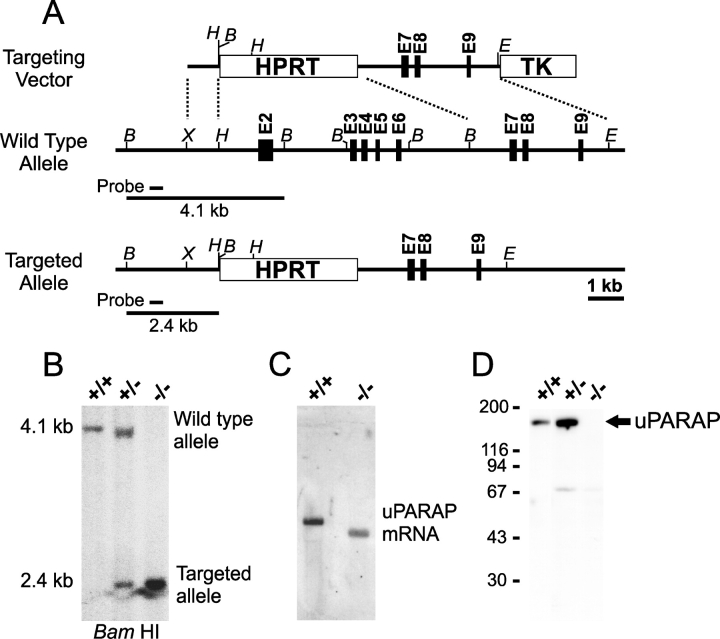
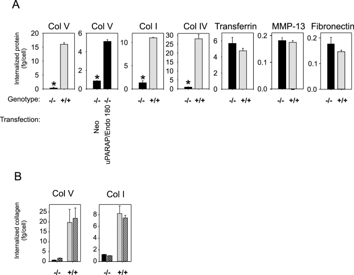
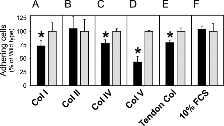
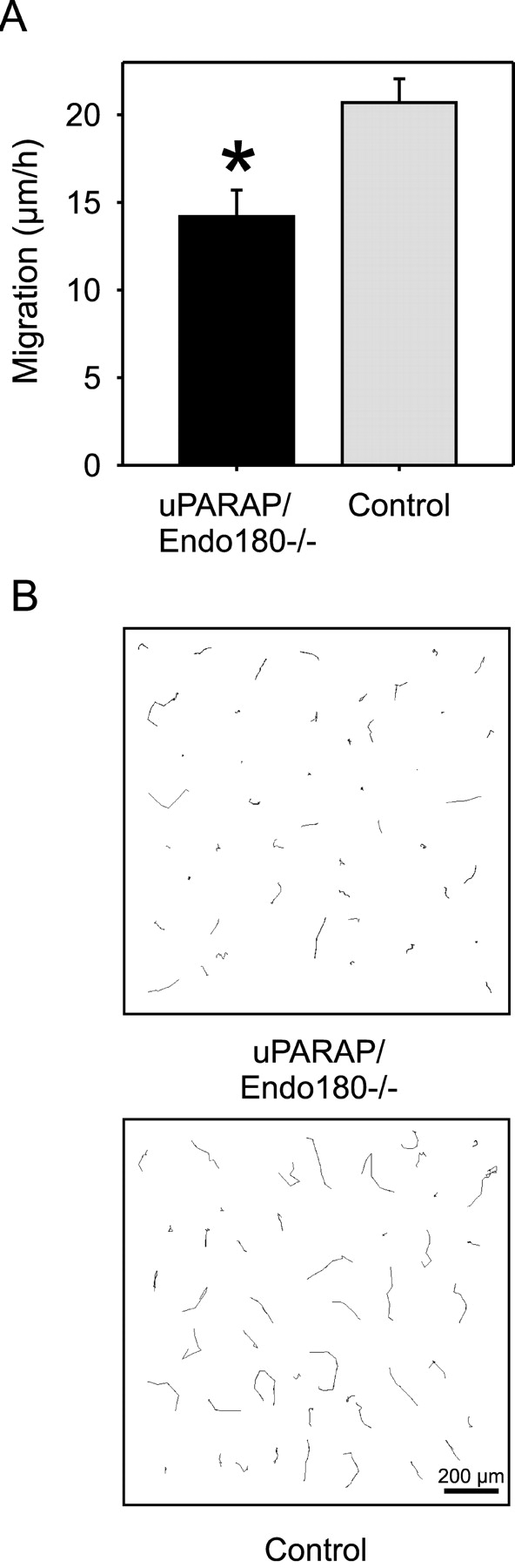
The uptake and lysosomal degradation of collagen by fibroblasts constitute a major pathway in the turnover of connective tissue. However, the molecular mechanisms governing this pathway are poorly understood. Here, we show that the urokinase plasminogen activator receptor-associated protein (uPARAP)/Endo180, a novel mesenchymally expressed member of the macrophage mannose receptor family of endocytic receptors, is a key player in this process. Fibroblasts from mice with a targeted deletion in the uPARAP/Endo180 gene displayed a near to complete abrogation of collagen endocytosis. Furthermore, these cells had diminished initial adhesion to a range of different collagens, as well as impaired migration on fibrillar collagen. These studies identify a central function of uPARAP/Endo180 in cellular collagen interactions.


Figures

Figure 1.
Figure 1.
Generation of uPARAP/Endo180-targeted mice. (A) Diagram of the targeting strategy showing the structure of the uPARAP/Endo180-targeting vector (top), wild-type uPARAP/Endo180 allele (middle), and targeted uPARAP/Endo180 allele (bottom). Exons 2–6 of the uPARAP/Endo180 gene (encoding the cysteine-rich, the FN-II, and the first carbohydrate recognition domains) were replaced by the HPRT selection cassette. Exons are indicated with boxes and introns with a solid line. B, BamHI; E, EcoRI; H, HindIII; X, XhoI. (B) Southern blot of BamHI-digested mouse tail DNA from mice genotyped by PCR as uPARAP/Endo180+/+ (left lane), uPARAP/Endo180+/− (middle lane), and uPARAP/Endo180−/− (right lane). The hybridization probe (solid line below the wild-type allele in A) was located upstream of the targeted area. The expected DNA fragments of the wild-type (4.1 kb) and targeted (2.4 kb) alleles that hybridize to the probe are indicated with solid lines in A. (C) Northern blot of total RNA isolated from cultured fibroblasts. RNA from fibroblasts of mice genotyped as uPARAP/Endo180+/+ (left lane) and uPARAP/Endo180−/− (right lane) was hybridized with a cDNA probe complementary to the 3′ end of the uPARAP/Endo180 mRNA. (D) Western blot of lysates of cultured dermal fibroblasts from neonates genotyped as uPARAP/Endo180+/+ (left lane), uPARAP/Endo180+/− (middle lane), and uPARAP/Endo180−/− (right lane). The blot was probed with a murine anti–uPARAP/Endo180 peptide antibody prepared as described in Materials and methods. The positions of molecular mass markers (kD) are indicated left.
Figure 2.
Figure 2.
uPARAP/Endo180 is required for collagen internalization. (A) Representative examples of the ability of dermal fibroblasts from uPARAP/Endo180−/− (black bars) or littermate uPARAP/Endo180+/+ (gray bars) neonates to internalize the 125I-labeled ligands indicated above each panel. Col I, IV, and V designate collagen subtypes. In panel 2, uPARAP/Endo180−/− fibroblasts were transiently transfected with a uPARAP/Endo180 expression plasmid, or with a neomycin resistance gene control expression plasmid, as indicated. The transfection efficiency was ∼10%. The data are expressed as the total amount of 125I-labeled ligand internalized per cell within a 3-h period at 37°C. Error bars indicate SDs. Asterisk in Col V, P < 0.000001; Col V in transfected cells, P < 0.000003; Col I, P < 0.00000001; and Col IV, P < 0.00005. (B) Cells with the genotype indicated were preincubated in the presence (hatched columns) or absence (unhatched columns) of a blocking antibody against β1 integrins, and the internalization of collagen V and I was measured as above.
Figure 3.
Figure 3.
uPARAP/Endo180 promotes cell adhesion to collagen. Dermal fibroblasts from uPARAP/Endo180−/− (black bars) or littermate uPARAP/Endo180+/+ neonates (gray bars) were allowed to attach for 30 min at 37°C to tissue culture wells coated with collagens type I (A), type II (B), type IV (C), type V (D), total tendon collagen (E), or serum proteins from 10% FCS (F). The number of cells adhering to the different matrices was determined by an MTT assay and expressed as the percentage of adherent uPARAP/Endo180+/+ cells. Error bars indicate SDs of quadruplicate determinations. The data are representative of results obtained with three different sets of uPARAP/Endo180−/− and littermate control fibroblasts. Asterisk in A, P < 0.03; C, P < 0.002; D, P < 0.0004; and E, P < 0.002.
Figure 4.
Figure 4.
uPARAP/Endo180 promotes cell migration on collagen. (A) The migration of dermal fibroblasts from uPARAP/Endo180−/− (black bar) or littermate control neonates (gray bar) on total tendon collagen was tracked for 6 h by time-lapse video microscopy. Data from individual cells of each genotype were collected in parallel recordings, and the rate of migration was calculated by pooling four independent sets of experiments, each showing similar results, that included cells from a total of four different uPARAP/Endo180−/− mice and five littermate control mice. A total of 52 uPARAP/Endo180−/− cells and 82 control cells were included in the measurements. Error bars indicate the standard error of the migration rates. Asterisk, P < 0.0016. (B) Migration paths on tendon collagen from 46 individual uPARAP/Endo180−/− and 46 individual control fibroblasts selected randomly. The cells were tracked for 6 h by time-lapse microscopy. Size bar is indicated in the lower right corner of the control cell panel.

References

    1. Ancian, P., G. Lambeau, and M. Lazdunski. 1995. Multifunctional activity of the extracellular domain of the M-type (180 kDa) membrane receptor for secretory phospholipases A2. Biochemistry. 34:13146–13151. - PubMed
    1. Bailey, L., D. Wienke, M. Howard, V. Knauper, C.M. Isacke, and G. Murphy. 2002. Investigation of the role of Endo180/urokinase-type plasminogen activator receptor-associated protein as a collagenase 3 (matrix metalloproteinase 13) receptor. Biochem. J. 363:67–72. - PMC - PubMed
    1. Barmina, O.Y., H.W. Walling, G.J. Fiacco, J.M. Freije, C. Lopez-Otin, J.J. Jeffrey, and N.C. Partridge. 1999. Collagenase-3 binds to a specific receptor and requires the low density lipoprotein receptor-related protein for internalization. J. Biol. Chem. 274:30087–30093. - PubMed
    1. Bateman, A., E. Birney, R. Durbin, S.R. Eddy, R.D. Finn, and E.L. Sonnhammer. 1999. Pfam 3.1: 1313 multiple alignments and profile HMMs match the majority of proteins. Nucleic Acids Res. 27:260–262. - PMC - PubMed
    1. Behrendt, N., E. Rønne, and K. Danø. 1996. Domain interplay in the urokinase receptor. Requirement for the third domain in high affinity ligand binding and demonstration of ligand contact sites in distinct receptor domains. J. Biol. Chem. 271:22885–22894. - PubMed

Publication types

MeSH terms