

Regulation of myotubularin-related (MTMR)2 phosphatidylinositol phosphatase by MTMR5, a catalytically inactive phosphatase
- PMID: 12668758
- PMCID: PMC153583
- DOI: 10.1073/pnas.0431052100
Regulation of myotubularin-related (MTMR)2 phosphatidylinositol phosphatase by MTMR5, a catalytically inactive phosphatase
Abstract
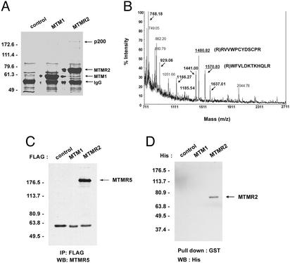
The myotubularin (MTM) family constitutes one of the most highly conserved protein-tyrosine phosphatase subfamilies in eukaryotes. MTM1, the archetypal member of this family, is mutated in X-linked myotubular myopathy, whereas mutations in the MTM-related (MTMR)2 gene cause the type 4B1 Charcot-Marie-Tooth disease, a severe hereditary motor and sensory neuropathy. In this study, we identified a protein that specifically interacts with MTMR2 but not MTM1. The interacting protein was shown by mass spectrometry to be MTMR5, a catalytically inactive member of the MTM family. We also demonstrate that MTMR2 interacts with MTMR5 via its coiled-coil domain and that mutations in the coiled-coil domain of either MTMR2 or MTMR5 abrogate this interaction. Through this interaction, MTMR5 increases the enzymatic activity of MTMR2 and dictates its subcellular localization. This article demonstrates an active MTM member being regulated by an inactive family member.
Figures


Similar articles
-
Membrane association of myotubularin-related protein 2 is mediated by a pleckstrin homology-GRAM domain and a coiled-coil dimerization module.Proc Natl Acad Sci U S A. 2003 Oct 14;100(21):12177-82. doi: 10.1073/pnas.2132732100. Epub 2003 Oct 6. Proc Natl Acad Sci U S A. 2003. PMID: 14530412 Free PMC article.
-
Loss of phosphatase activity in myotubularin-related protein 2 is associated with Charcot-Marie-Tooth disease type 4B1.Hum Mol Genet. 2002 Jun 15;11(13):1569-79. doi: 10.1093/hmg/11.13.1569. Hum Mol Genet. 2002. PMID: 12045210
-
Multi-level regulation of myotubularin-related protein-2 phosphatase activity by myotubularin-related protein-13/set-binding factor-2.Hum Mol Genet. 2006 Feb 15;15(4):569-79. doi: 10.1093/hmg/ddi473. Epub 2006 Jan 6. Hum Mol Genet. 2006. PMID: 16399794
-
Charcot-Marie-Tooth type 4B demyelinating neuropathy: deciphering the role of MTMR phosphatases.Expert Rev Mol Med. 2007 Sep 20;9(25):1-16. doi: 10.1017/S1462399407000439. Expert Rev Mol Med. 2007. PMID: 17880751 Review.
-
The structure and regulation of myotubularin phosphatases.Curr Opin Struct Biol. 2005 Dec;15(6):614-20. doi: 10.1016/j.sbi.2005.10.016. Epub 2005 Nov 9. Curr Opin Struct Biol. 2005. PMID: 16289848 Review.
Cited by
-
Defective membrane remodeling in neuromuscular diseases: insights from animal models.PLoS Genet. 2012;8(4):e1002595. doi: 10.1371/journal.pgen.1002595. Epub 2012 Apr 5. PLoS Genet. 2012. PMID: 22496665 Free PMC article. Review.
-
Expression patterns and the roles of phosphatidylinositol phosphatases in testis†.Biol Reprod. 2022 Oct 11;107(4):902-915. doi: 10.1093/biolre/ioac132. Biol Reprod. 2022. PMID: 35766372 Free PMC article.
-
Endosomal targeting of the phosphoinositide 3-phosphatase MTMR2 is regulated by an N-terminal phosphorylation site.J Biol Chem. 2011 May 6;286(18):15841-53. doi: 10.1074/jbc.M110.209122. Epub 2011 Mar 3. J Biol Chem. 2011. PMID: 21372139 Free PMC article.
-
The role of myotubularin-related phosphatases in the control of autophagy and programmed cell death.Adv Biol Regul. 2012 Jan;52(1):282-9. doi: 10.1016/j.advenzreg.2011.10.001. Adv Biol Regul. 2012. PMID: 22056831 Free PMC article. Review. No abstract available.
-
Characterization of a novel zebrafish model of MTMR5-associated Charcot-Marie-Tooth disease type 4B3.Brain Commun. 2025 Feb 18;7(2):fcaf077. doi: 10.1093/braincomms/fcaf077. eCollection 2025. Brain Commun. 2025. PMID: 40066109 Free PMC article.
References
-
- Laporte J, Blondeau F, Buj-Bello A, Mandel J-L. Trends Genet. 2001;17:221–228. - PubMed
-
- Maehama T, Taylor G S, Dixon J E. Annu Rev Biochem. 2001;70:247–279. - PubMed
-
- Wishart M J, Taylor G S, Slama J T, Dixon J E. Curr Opin Cell Biol. 2001;13:172–181. - PubMed
-
- Denu J M, Dixon J E. Curr Opin Chem Biol. 1998;2:633–641. - PubMed
-
- Cui X, De Vivo I, Slany R, Miyamoto A, Firestein R, Cleary M L. Nat Genet. 1998;18:331–337. - PubMed
Publication types
MeSH terms
Substances
LinkOut - more resources
Full Text Sources
Other Literature Sources
Molecular Biology Databases
