. 2003 Apr 15;100(8):4610-5.
doi: 10.1073/pnas.0835895100. Epub 2003 Mar 31.

Stabilization of beta-catenin by a Wnt-independent mechanism regulates cardiomyocyte growth

Affiliations

Stabilization of beta-catenin by a Wnt-independent mechanism regulates cardiomyocyte growth

Syed Haq et al. Proc Natl Acad Sci U S A. .

Abstract

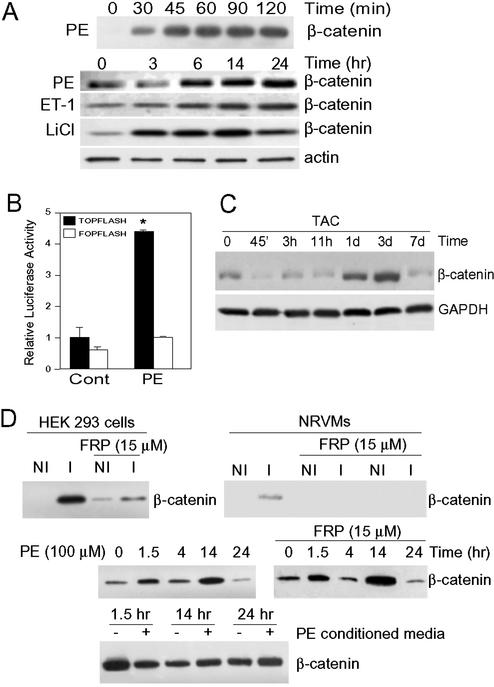
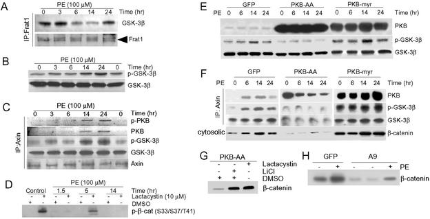
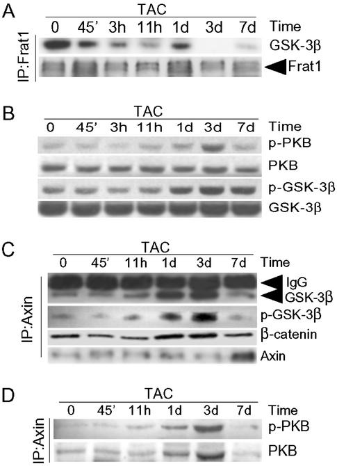
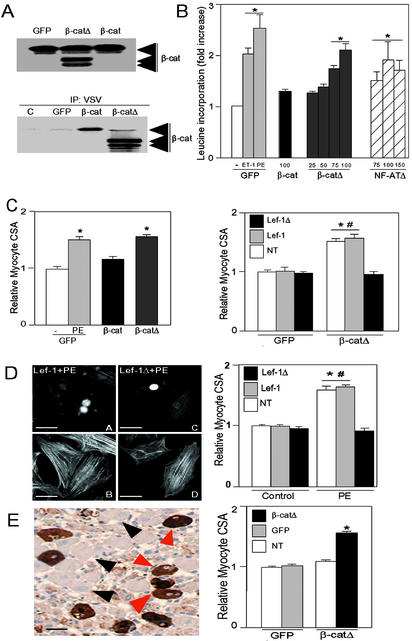
beta-Catenin is a transcriptional activator that regulates embryonic development as part of the Wnt pathway and also plays a role in tumorigenesis. The mechanisms leading to Wnt-induced stabilization of beta-catenin, which results in its translocation to the nucleus and activation of transcription, have been an area of intense interest. However, it is not clear whether stimuli other than Wnts can lead to important stabilization of beta-catenin and, if so, what factors mediate that stabilization and what biologic processes might be regulated. Herein we report that beta-catenin is stabilized in cardiomyocytes after these cells have been exposed to hypertrophic stimuli in culture or in vivo. The mechanism by which beta-catenin is stabilized is distinctly different from that used by Wnt signaling. Although, as with Wnt signaling, inhibition of glycogen synthase kinase-3 remains central to hypertrophic stimulus-induced stabilization of beta-catenin, the mechanism by which this occurs involves the recruitment of activated PKB to the beta-catenin-degradation complex. PKB stabilizes the complex and phosphorylates glycogen synthase kinase-3 within the complex, inhibiting its activity directed at beta-catenin. Finally, we demonstrate via adenoviral gene transfer that beta-catenin is both sufficient to induce growth in cardiomyocytes in culture and in vivo and necessary for hypertrophic stimulus-induced growth. Thus, in these terminally differentiated cells, beta-catenin is stabilized by hypertrophic stimuli acting via heterotrimeric G protein-coupled receptors. The stabilization occurs via a unique Wnt-independent mechanism and results in cellular growth.


Figures

Figure 1
Figure 1
Hypertrophic stimuli induce β-catenin accumulation in vitro and in vivo. (A) NRVMs were treated with PE (100 μM), endothelin-1 (100 nM), or, as a positive control, with the GSK-3 inhibitor LiCl (10 mM), for the times indicated. Cytosolic fractions were blotted with anti-β-catenin antibody. Antiactin blot confirms equivalent protein loading. (B) NRVMs were transfected with pTOPFlash or pFOPFlash and 24 h later were stimulated with PE or vehicle (Cont) for an additional 24 h before luciferase assay. Luciferase activity of vehicle-treated, pTOPFlash-transfected cells was set at 1, and other results are expressed as fold activation relative to that value (n = 5 independent experiments, done in triplicate; *, P < 0.01 vs. all other values). (C) Rats were subjected to TAC or sham surgery (control) for the times indicated. Lysates of cytosolic fractions were blotted with anti-β-catenin antibody. Anti-GAPDH blot confirms equivalent loading. Shams had no increase in β-catenin throughout the 7-d protocol (data not shown). (D Top), NRVMs and HEK293 cells were treated for 2 h with media from S2 cells that had not been induced to secrete Wg (NI) or were induced (I) by the addition of CdCl2 (see Methods). A separate group of cells was pretreated with Frizzled-related protein-1 (FRP, 15 μM) for 1 h before the addition of NI or I S2 media for 2 h. Cytosolic fractions were blotted with anti-β-catenin antibody. (Middle) NRVMs were incubated with PE (100 μM), with or without Frizzled-related protein-1 pretreatment for 1 h, for the times indicated. Cytosolic fractions were blotted with anti-β-catenin antibody. (Bottom) NRVMs were treated with or without PE for 14 h, after which the conditioned media were collected and incubated for the times indicated with fresh cultures of NRVMs. Cytosolic fractions were blotted with anti-β-catenin antibody.
Figure 2
Figure 2
Mechanisms of β-catenin stabilization in cardiomyocytes. NRVMs were stimulated with PE (100 μM) for the times indicated. (A) Lysates were subjected to immunoprecipitation with anti-Frat1 antibody followed by blotting with antibodies to GSK-3β or Frat1. (B) Lysates were blotted with antibodies to Ser-9-phosphorylated GSK-3β (p-GSK-3β) or to GSK-3β. (C) Lysates were subjected to immunoprecipitation with anti-Axin antibody followed by blotting with antibodies to p-GSK-3β, GSK-3β, Axin, or with an antibody recognizing Ser-473-phosphorylated (active) PKB (p-PKB) or total PKB. (D) NRVMs were pretreated with either DMSO or lactacystin (10 μM) for 1 h before stimulation with PE for the times indicated. Cytosolic fractions were prepared and then blotted with anti-phospho-specific Ser-33/37/Thr-41 β-catenin antibody. (E and F) NRVMs were transduced with AdPKB-AA, AdPKB-myr, or AdGFP (control) for 48 h by using a multiplicity of infection of 25 plaque-forming units per cell and then stimulated with PE for the times indicated. Cell lysates were blotted with antibodies to PKB, p-GSK-3β, and GSK-3β (E), or lysates were immunoprecipitated with anti-Axin antibody followed by blotting with antibodies to PKB, p-GSK-3β, GSK-3β (F). Cytosolic fractions were blotted with anti-β-catenin antibody (Bottom). (G) NRVMs were transduced with AdPKB-AA and 48 h later were incubated for 2 h with LiCl, lactacystin, or DMSO, as indicated. Cytosolic fractions were blotted with anti-β-catenin antibody. (H) NRVMs were transduced with either AdGSK-3β(S9A) or AdGFP for 24 h and then stimulated with PE or vehicle (-) for 14 h. Cytosolic fractions were blotted with anti-β-catenin antibody.
Figure 3
Figure 3
Mechanisms of hypertrophic stress-induced stabilization of β-catenin in vivo. Rats were subjected to TAC for the times indicated. (A) Myocardial lysates were immunoprecipitated with anti-Frat1 antibody, followed by blotting with anti-GSK-3β and anti-Frat1 antibodies. (B) Myocardial lysates were blotted with antibodies to p-PKB, PKB, p-GSK-3β, or GSK-3β. (C and D) Myocardial lysates were immunoprecipitated with an anti-Axin antibody followed by blotting with antibodies to GSK-3β, p-GSK-3β, and β-catenin (C) or with anti-p-PKB or anti-PKB (D).
Figure 4
Figure 4
β-Catenin is sufficient and necessary for cardiomyocyte hypertrophy. (A) NRVMs were transduced with AdGFP (GFP), Adβ-catenin (β-cat), or Adβ-cateninΔ (β-catΔ), each with a multiplicity of infection of 100 plaque-forming units per cell. Lysates were blotted with anti-β-catenin antibody (Upper) or were subjected to immunoprecipitation with anti-vesicular stomatitis virus antibody followed by blotting with anti-β-catenin antibody (Lower). (B) NRVMs were transduced with AdGFP, Adβ-catenin, Adβ-cateninΔ, or AdNF-ATΔ at the multiplicities of infection noted. Incorporation of [3H]leucine was determined 48 h later. As a reference, [3H]leucine incorporation was determined in AdGFP-transduced cells stimulated with endothelin-1 or PE for 48 h. Values for AdGFP-transduced cells treated with vehicle were normalized to 1 (n = 5–6 experiments, each done in triplicate). *, P < 0.01 vs. AdGFP-transduced cells treated with vehicle. (C Left), β-CateninΔ induces an increase in cardiomyocyte size in vitro. NRVMs on coverslips were transduced with AdGFP, Adβ-catenin, or Adβ-cateninΔ at a multiplicity of infection of 100 plaque-forming units per cell. AdGFP-transduced cells were stimulated with PE or vehicle (-) for 48 h. Transduced cells were identified by GFP positivity, and cardiomyocyte CSA was determined on images of anti-α-actinin-stained myocytes (n = 4 experiments; ≥75 myocytes measured per experiment). *, P < 0.01 vs. AdGFP-transduced cells treated with vehicle. (Right) Inhibition of β-cateninΔ-induced hypertrophy by Lef-1Δβ-catenin. NRVMs were transduced with AdGFP or Adβ-cateninΔ and then were transfected with plasmids encoding Myc epitope-tagged wild-type Lef-1 or Lef-1Δβ-catenin. Cardiomyocyte CSA was determined 48 h later in cells successfully transfected with either Lef-1 or Lef-1Δβ-catenin (Lef-1Δ) and in cells on the same coverslips that had undergone the transfection protocol but were not successfully transfected (NT; n = 4 experiments; ≥75 myocytes measured per experiment). #, P < 0.01 vs. CSA of Adβ-catΔ-transduced cells that were transfected with Lef-1Δβ-cat. *, P < 0.01 vs. CSA of all AdGFP-transduced cells. (D) Inhibition of PE-induced hypertrophy by Lef-1Δβ-catenin. (Left) NRVMs were transfected with plasmids encoding Myc-tagged wild-type Lef-1 (A and B) or Myc-tagged Lef-1Δβ-cat (C and D). Twenty-four hours later, cells were stimulated with PE. Cardiomyocyte CSA was determined 48 h later. (A and C) Immunostained with anti-Myc tag to identify transfected cells. (B and D) The same fields stained with FITC-conjugated phalloidin. (Right) Graph compares CSA of cells successfully transfected with either Lef-1 or Lef-1Δβ-cat with cells that were not transfected (NT), after stimulation with PE or vehicle (control). Data are from four experiments; ≥75 myocytes measured per experiment. *, P < 0.01 vs. CSA of all controls. #, P < 0.01 vs. CSA of Lef-1Δβ-cat-transfected cells. (Bar, 20 μm.) (E) β-CateninΔ increased cardiomyocyte size in vivo. (Left) AdGFP or Adβ-cateninΔ were injected directly into the left ventricular myocardium of rats. Eight days, later cardiomyocyte CSA was determined on GFP-positive myocytes infected with Adβ-cateninΔ (red arrows) and adjacent nontransduced myocytes (black arrows). (Bar, 20 μm.) (Right) Graph compares CSA of cells successfully transduced with Adβ-cateninΔ, AdGFP, or cells that were not transduced (NT; n = 4 hearts per treatment group; ≥50 myocytes measured per heart). *, P < 0.01 vs. AdGFP transduced.

Similar articles

Cited by

References

    1. He X, Saint-Jeannet J P, Woodgett J R, Varmus H E, Dawid I B. Nature. 1995;374:617–622. - PubMed
    1. Haq S, Choukroun G, Kang Z B, Lee K-H, Ranu H, Matsui T, Rosenzweig A, Alessandrini A, Molkentin J D, Woodgett J, et al. J Cell Biol. 2000;151:117–129. - PMC - PubMed
    1. Morisco C, Zebrowski D, Condorelli G, Tsichlis P, Vatner S F, Sadoshima J. J Biol Chem. 2000;275:14466–14475. - PubMed
    1. Badorff C, Ruetten H, Mueller S, Stahmer M, Gehring D, Jung F, Ihling C, Zeiher A M, Dimmeler S. J Clin Invest. 2002;3:373–381. - PMC - PubMed
    1. Antos C L, McKinsey T A, Frey N, Kutschke W, McAnally J, Shelton J M, Richardson J A, Hill J A, Olson E N. Proc Natl Acad Sci USA. 2002;99:907–912. - PMC - PubMed

Publication types

MeSH terms

LinkOut - more resources