

Mutational analysis of Ctnnb1 and Apc in tumors from rats given 1,2-dimethylhydrazine or 2-amino-3-methylimidazo[4,5-f]quinoline: mutational 'hotspots' and the relative expression of beta-catenin and c-jun
- PMID: 12669311
- PMCID: PMC2279233
- DOI: 10.1002/mc.10112
Mutational analysis of Ctnnb1 and Apc in tumors from rats given 1,2-dimethylhydrazine or 2-amino-3-methylimidazo[4,5-f]quinoline: mutational 'hotspots' and the relative expression of beta-catenin and c-jun
Abstract
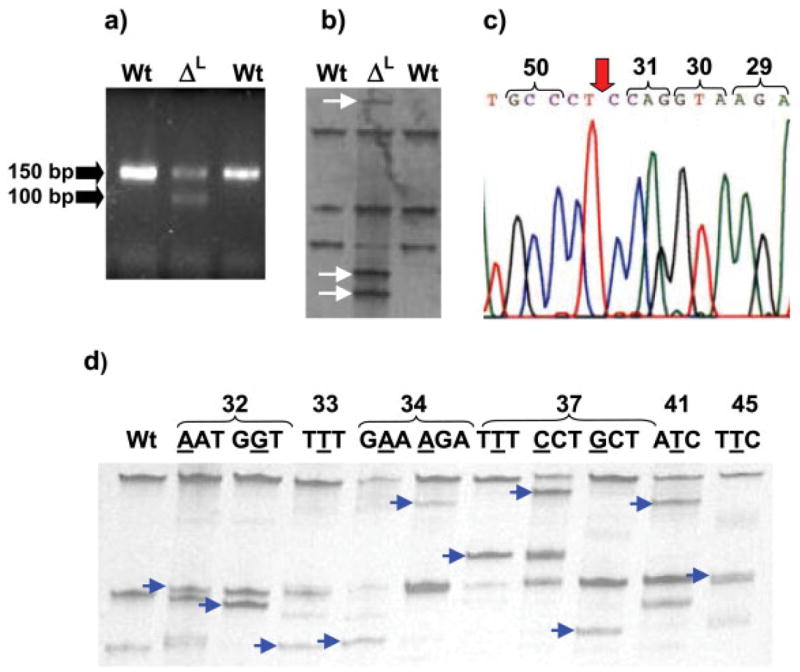
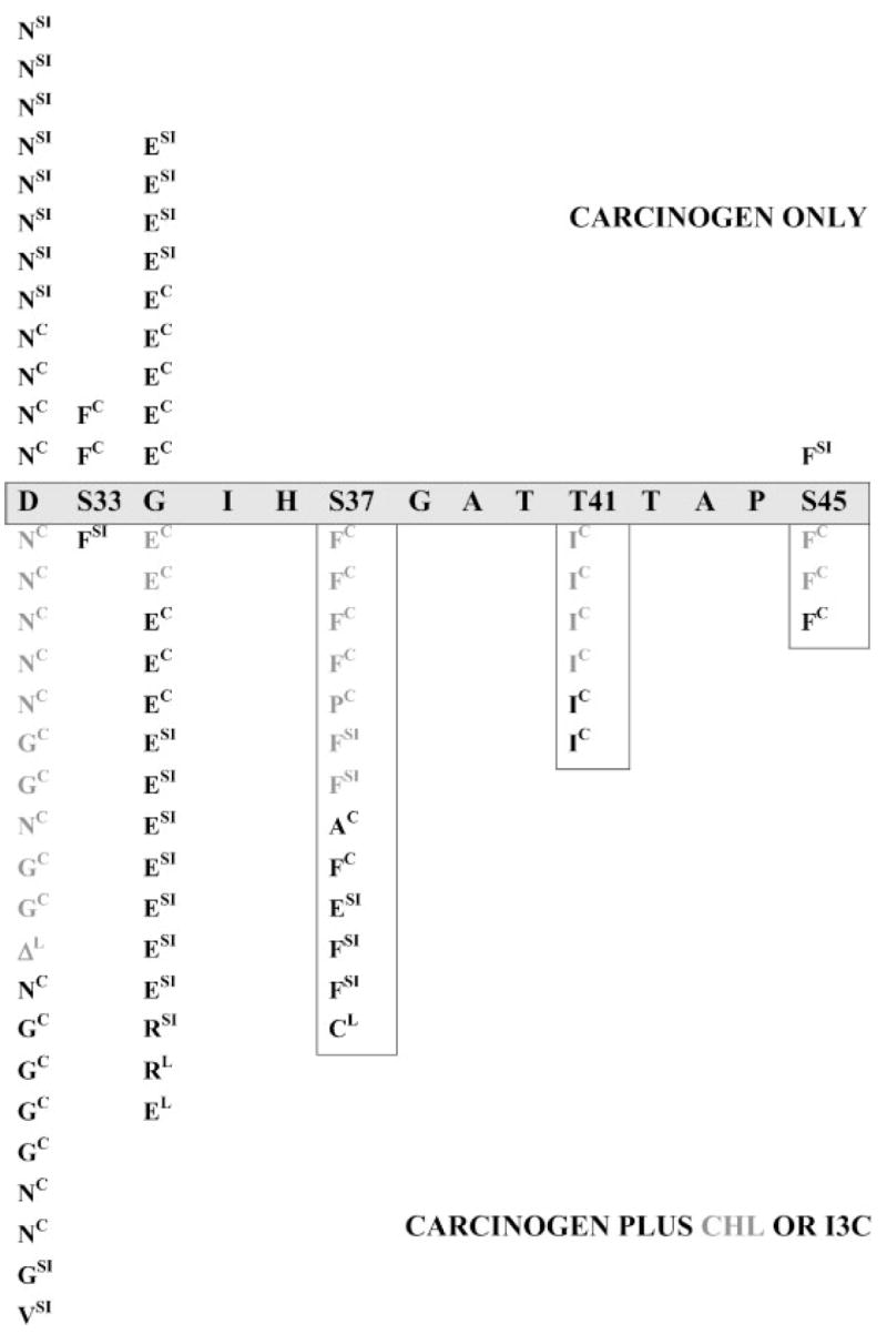
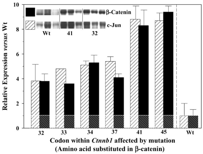
There is growing interest in beta-catenin and its role in various human cancers. We recently reported that 2-amino-3-methylimidazo[4,5-f]quinoline (IQ)- and 1,2-dimethylhydrazine (DMH)-induced colon tumors in the rat contain mutations in Ctnnb1, the gene for beta-catenin, but the mutation spectrum was influenced by postinitiation exposure to chlorophyllin (CHL) and indole-3-carbinol (I3C) [Blum et al., Carcinogenesis 2001;22:315-320]. The present paper describes a follow-up study in which all of the target organs for IQ- and DMH-induced tumorigenesis were screened; Ctnnb1 mutations were found in 44 of 119 DMH-induced colon tumors, six of 13 IQ-induced colon tumors, 28 of 81 DMH-induced small intestine tumors, none of five IQ-induced small intestine tumors, four of 106 IQ-induced liver tumors, none of 14 DMH-induced Zymbal's gland tumors, none of 24 IQ-induced Zymbal's gland tumors, and none of 29 IQ-induced skin tumors. In tumors from rats given carcinogen alone, or carcinogen plus CHL or I3C, Ctnnb1 mutations frequently substituted amino acids adjacent to Ser33, a critical Ser/Thr residue in the glycogen synthase kinase-3beta regulatory domain of beta-catenin. However, substitution of critical Ser/Thr residues themselves was detected in only three of 24 (12.5%) of the tumors from rats given carcinogen alone, compared with 23 of 58 (40%) of the tumors from rats given carcinogen and treated postinitiation with I3C or CHL (P < 0.02). More than 50 of the colon tumors with wild-type beta-catenin were examined further for their Apc status; the overall frequency of Apc mutations was <10%, and these genetic changes occurred exclusively in the 'Mutation Cluster Region' of Apc. A subset of colon tumors also was examined for expression of beta-catenin and c-jun; these proteins were overexpressed in all tumors containing Ctnnb1 mutations, but the expression was highest in tumors with Ctnnb1 mutations affecting Thr41 and Ser45 residues in the glycogen synthase kinase-3beta region of beta-catenin. Thus, Ctnnb1 mutations occurred more frequently than Apc mutations in colon and small intestine tumors of the rat, and certain mutations upregulated beta-catenin/T-cell factor target genes more effectively than others, perhaps influencing the response to phytochemicals administered postinitiation.
Copyright 2003 Wiley-Liss, Inc.
Figures
Similar articles
-
beta-Catenin mutation in rat colon tumors initiated by 1,2-dimethylhydrazine and 2-amino-3-methylimidazo[4,5-f]quinoline, and the effect of post-initiation treatment with chlorophyllin and indole-3-carbinol.Carcinogenesis. 2001 Feb;22(2):315-20. doi: 10.1093/carcin/22.2.315. Carcinogenesis. 2001. PMID: 11181454
-
Promotion versus suppression of rat colon carcinogenesis by chlorophyllin and chlorophyll: modulation of apoptosis, cell proliferation, and beta-catenin/Tcf signaling.Mutat Res. 2003 Feb-Mar;523-524:217-23. doi: 10.1016/s0027-5107(02)00338-x. Mutat Res. 2003. PMID: 12628520
-
Tumors from rats given 1,2-dimethylhydrazine plus chlorophyllin or indole-3-carbinol contain transcriptional changes in beta-catenin that are independent of beta-catenin mutation status.Mutat Res. 2006 Oct 10;601(1-2):11-8. doi: 10.1016/j.mrfmmm.2006.05.026. Epub 2006 Jul 24. Mutat Res. 2006. PMID: 16860348 Free PMC article.
-
Genetic changes induced by heterocyclic amines.Mutat Res. 1997 May 12;376(1-2):161-7. doi: 10.1016/s0027-5107(97)00039-0. Mutat Res. 1997. PMID: 9202752 Review.
-
Gene mutations and altered gene expression in azoxymethane-induced colon carcinogenesis in rodents.Cancer Sci. 2004 Jun;95(6):475-80. doi: 10.1111/j.1349-7006.2004.tb03235.x. Cancer Sci. 2004. PMID: 15182426 Free PMC article. Review.
Cited by
-
Hepatoprotective effect of Matricaria chamomilla aqueous extract against 1,2-Dimethylhydrazine-induced carcinogenic hepatic damage in mice.Heliyon. 2020 Jun 1;6(6):e04082. doi: 10.1016/j.heliyon.2020.e04082. eCollection 2020 Jun. Heliyon. 2020. PMID: 32509999 Free PMC article.
-
Bcl-2 overexpression in PhIP-induced colon tumors: cloning of the rat Bcl-2 promoter and characterization of a pathway involving beta-catenin, c-Myc and E2F1.Oncogene. 2007 Sep 13;26(42):6194-202. doi: 10.1038/sj.onc.1210438. Epub 2007 Apr 2. Oncogene. 2007. PMID: 17404573 Free PMC article.
-
Protective versus promotional effects of white tea and caffeine on PhIP-induced tumorigenesis and beta-catenin expression in the rat.Carcinogenesis. 2008 Apr;29(4):834-9. doi: 10.1093/carcin/bgn051. Epub 2008 Feb 17. Carcinogenesis. 2008. PMID: 18283038 Free PMC article.
-
Involvement of mutation-based inhibition of beta-catenin phosphorylation at Ser33 in the malignant progression of lung (pre)neoplastic lesions induced by N-nitrosobis(2-hydroxypropyl)amine in male Fischer 344 rats.Lung. 2007 Sep-Oct;185(5):271-278. doi: 10.1007/s00408-007-9017-y. Epub 2007 Jul 18. Lung. 2007. PMID: 17639448
-
Mouse models for the study of colon carcinogenesis.Carcinogenesis. 2009 Feb;30(2):183-96. doi: 10.1093/carcin/bgn267. Epub 2008 Nov 26. Carcinogenesis. 2009. PMID: 19037092 Free PMC article. Review.
References
-
- Polakis P. Wnt signaling and cancer. Genes Dev. 2000;14:1837–1851. - PubMed
-
- Oving IM, Clevers HC. Molecular causes of colon cancer. Eur J Clin Invest. 2002;32:448–457. - PubMed
-
- Clements WM, Wang J, Sarnaik A, et al. β-Catenin mutation is a frequent cause of Wnt pathway activation in gastric cancer. Cancer Res. 2002;62:3503–3506. - PubMed
-
- Kinzler KW, Vogelstein B. Cancer-susceptibility genes. Gatekeepers and caretakers. Nature. 1997;386:761–763. - PubMed
Publication types
MeSH terms
Substances
Grants and funding
LinkOut - more resources
Full Text Sources
Medical
Miscellaneous
