. 2003 Apr 1;17(7):838-43.
doi: 10.1101/gad.1064803.

In vivo analysis of a developmental circuit for direct transcriptional activation and repression in the same cell by a Runx protein

Affiliations

In vivo analysis of a developmental circuit for direct transcriptional activation and repression in the same cell by a Runx protein

Jude Canon et al. Genes Dev. .

Abstract

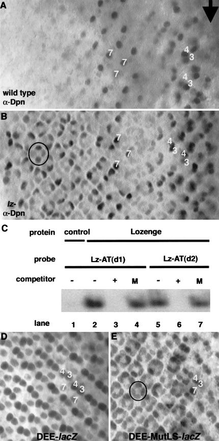
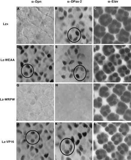
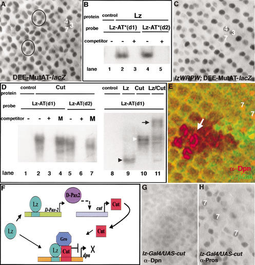
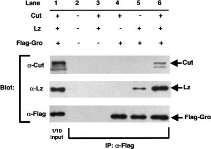
Runx proteins have been implicated in acute myeloid leukemia, cleidocranial dysplasia, and stomach cancer. These proteins control key developmental processes in which they function as both transcriptional activators and repressors. How these opposing regulatory modes can be accomplished in the in vivo context of a cell has not been clear. In this study we use the developing cone cell in the Drosophila visual system to elucidate the mechanism of positive and negative regulation by the Runx protein Lozenge (Lz). We describe a regulatory circuit in which Lz causes transcriptional activation of the homeodomain protein Cut, which can then stabilize a Lz repressor complex in the same cell. Whether a gene is activated or repressed is determined by whether the Lz activator or the repressor complex binds to its upstream sequence. This study provides a mechanistic basis for the dual function of Runx proteins that is likely to be conserved in mammalian systems.


Figures

Figure 1
Figure 1
Lz directly represses dpn transcription in cone cells. (A) In wild type, Dpn is expressed in R3/R4 cells for four to five columns posterior to the furrow (arrow), then in R7 cells for five to six columns. (B) In lz mutants, Dpn is also ectopically activated in cone cells (one cluster circled). (C) Lz binds to the two Runx consensus binding sites in the dpn enhancer. Nuclear extracts were made from S2 cells transfected with vector alone (control) or a Lz-expressing vector. For the sequences of all probes used, see Table 1. Lz binds both sites (lanes 2,5), and this binding is specific as it is competed away with cold competitor oligo (+; lanes 3,6), but not by oligo containing a mutation (M) in the Runx-binding site (lanes 4,7). The portion of the gel shown represents the only shifted bands above the free probe. (D) The dpn eye enhancer (DEE) drives expression of a lacZ reporter in R3/R4 and R7, like the wild-type Dpn expression pattern, except that the expression of β-Gal perdures to the back of the eye disc. (E) When the two Lz-binding sites (LBS) are mutated in the DEE, expression is also seen in cone cells (one cluster circled), indicating that Lz directly represses the DEE in those cells.
Figure 2
Figure 2
The C terminus of Lz, a Gro-interaction domain, influences the mode of Lz regulation. lz transgenes were tested for their ability to regulate Lz targets dpn and D-Pax2. Transgenes and antibodies are as indicated. Stainings are of eye discs expressing one copy of the transgene in a lz77a7/Y mutant background. Circles mark one cluster of cone cells. Wild-type Lz+ represses dpn (A), but activates D-Pax2 (B) in cone cells. Lz–WEAA is unable to repress dpn in cone cells (D), but can activate D-Pax2 expression (E). Lz–WRPW represses dpn in cone cells (G), but is unable to activate D-Pax2 (H). Therefore, Lz–WRPW functions as a dedicated repressor. Lz–VP16 cannot repress dpn in cone cells (J), but activates D-Pax2 (K), and therefore functions as a constitutive activator. Expression of the neuronal differentiation marker Elav is unchanged in all the above genetic backgrounds (C,F,I,L).
Figure 3
Figure 3
Cut binding to the AT-rich sequences adjacent to the Lz-binding sites is required for dpn repression. (A) Mutation of both AT-rich sequences in the DEE causes derepression of this enhancer, as lacZ reporter gene expression is now also seen in cone cells (two clusters circled; cf. with Fig. 1D). (B) Lz binding is not affected by mutations in the AT-rich sequences. Nuclear extracts were made from S2 cells transfected with vector alone (control) or with a Lz-expressing vector. For probe sequences, see Table 1. Lz binds to both of its binding sites (lanes 2,4) even when the adjacent AT-sequences are altered. This binding is specific as it is competed away with cold competitor (lanes 3,5). (C) Lz–WRPW is able to repress the DEE in cone cells despite the mutated AT-rich sites. The lacZ reporter gene is expressed in R3/R4 and R7, but not in the cone cells. (D) Cut binds to the AT-rich sequences in the dpn enhancer. Nuclear extracts were made from cells expressing proteins as indicated. Probes are defined in Table 1. Left and right panels represent different gels. (Left panel) Cut binds to the two AT-rich sequences in the DEE (lanes 2,5), and this binding is specific as it is competed away with cold oligo containing known Cut-binding sites (lanes 3,6), but not by cold oligo that contains a mutation (M) in the AT-rich sites (lanes 4,7). (Right panel) A probe containing a Lz-binding site and adjacent AT sequences from the dpn enhancer [Lz–AT(d1); Table 1] can bind Lz (black arrowhead, lane 9) and Cut (white arrowhead, lane 10). Extracts from cells transfected with both lz and cut cause a supershifted band (arrow, lane 11) indicating that Lz and Cut can together bind the same probe. (E) Dpn is ectopically expressed in cone cells in clones of cut mutant cells in the eye. Arrow indicates cone cells within the mutant clone (nongreen) expressing Dpn (red). The counter marker β-Gal (green) marks wild-type tissue. (F) Model for a developmental circuit for positive and negative regulation by Lz in cone cells. Lz directly activates D-Pax2, which is required for activation of cut. Cut protein then binds and forms a repressive complex with Lz and Gro that represses dpn in cone cells. (G) Misexpression of Cut in R7 cells using the lz–Gal4 driver causes repression of dpn in those cells. (H) The R7-cell-specific marker Prospero is unchanged in this genetic background.
Figure 4
Figure 4
Cut stabilizes an interaction between Lz and Gro. Nuclear extracts were made from S2 cells transfected with lz, cut, and Flag–gro as indicated, and used in immunoprecipitation experiments. Lane 1 shows blots of extracts (1/10 of input), and lanes 2–6 are blots of extracts immunoprecipitated with α-Flag antibody. Extracts in lane 2 are from cells transfected with vector alone. The control in lane 3 shows that α-Flag does not precipitate Lz or Cut in the absence of Gro. Also, Flag–Gro does not interact with Cut (lane 4). A small amount of Lz coimmunoprecipitates with Gro (lane 5), but this interaction is significantly increased in the presence of Cut (lane 6). The same results were obtained in the presence of DNA containing Lz- and Cut-binding sites (data not shown).

References

    1. Aronson BD, Fisher AL, Blechman K, Caudy M, Gergen JP. Groucho-dependent and -independent repression activities of Runt domain proteins. Mol Cell Biol. 1997;17:5581–5587. - PMC - PubMed
    1. Bier E, Vaessin H, Younger-Shepherd S, Jan LY, Jan YN. deadpan, an essential pan-neural gene in Drosophila, encodes a helix–loop–helix protein similar to the hairy gene product. Genes & Dev. 1992;6:2137–2151. - PubMed
    1. Blochlinger K, Bodmer R, Jack J, Jan LY, Jan YN. Primary structure and expression of a product from cut, a locus involved in specifying sensory organ identity in Drosophila. Nature. 1988;333:629–635. - PubMed
    1. Canon J, Banerjee U. Runt and Lozenge function in Drosophila development. Semin Cell Dev Biol. 2000;11:327–336. - PubMed
    1. Castilla LH, Wijmenga C, Wang Q, Stacy T, Speck NA, Eckhaus M, Marin-Padilla M, Collins FS, Wynshaw-Boris A, Liu PP. Failure of embryonic hematopoiesis and lethal hemorrhages in mouse embryos heterozygous for a knocked-in leukemia gene CBFB-MYH11. Cell. 1996;87:687–696. - PubMed

Publication types

MeSH terms

LinkOut - more resources