. 2003 Apr 1;17(7):912-25.
doi: 10.1101/gad.250603.

Transcriptional oscillation of lunatic fringe is essential for somitogenesis

Affiliations

Transcriptional oscillation of lunatic fringe is essential for somitogenesis

Katrin Serth et al. Genes Dev. .

Abstract

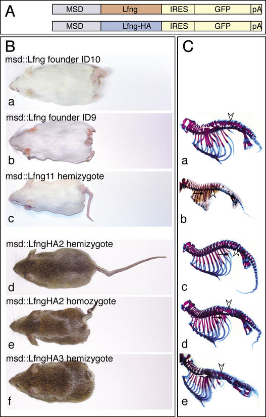
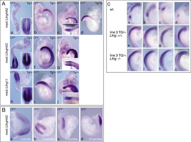
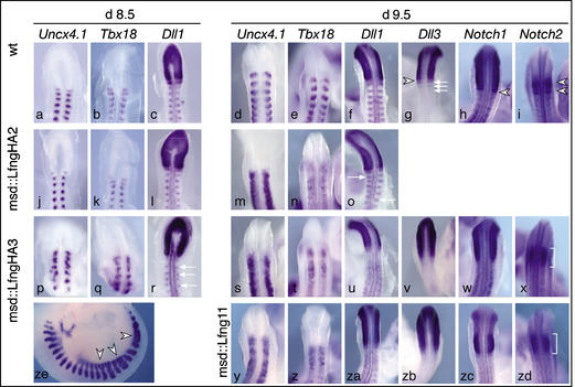
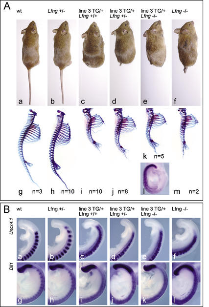
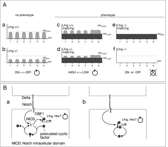
A molecular oscillator that controls the expression of cyclic genes such as lunatic fringe (Lfng) in the presomitic mesoderm has been shown to be coupled with somite formation in vertebrate embryos. To address the functional significance of oscillating Lfng expression, we have generated transgenic mice expressing Lfng constitutively in the presomitic mesoderm in addition to the intrinsic cyclic Lfng activity. These transgenic lines displayed defects of somite patterning and vertebral organization that were very similar to those of Lfng null mutants. Furthermore, constitutive expression of exogenous Lfng did not compensate for the complete loss of cyclic endogenous Lfng activity. Noncyclic exogenous Lfng expression did not abolish cyclic expression of endogenous Lfng in the posterior presomitic mesoderm (psm) but affected its expression pattern in the anterior psm. Similarly, dynamic expression of Hes7 was not abolished but abnormal expression patterns were obtained. Our data are consistent with a model in which alternations of Lfng activity between ON and OFF states in the presomitic mesoderm prior to somite segmentation are critical for proper somite patterning, and suggest that Notch signaling might not be the only determinant of cyclic gene expression in the presomitic mesoderm of mouse embryos.


Figures

Figure 1
Figure 1
Structure of transgenes and external and skeletal phenotype of transgenic mice. (A) Structure of the msd∷Lfng and msd∷LfngHA transgenes. msd refers to the portion of the Delta1 promoter directing heterologous gene expression into the paraxial mesoderm (Beckers et al. 2000). (B) Transgenic founder mice obtained with msd∷Lfng (panels a,b), a hemizygous msd∷Lfng11 mouse (panel c), hemizygous (panel d) and homozygous (panel e) transgenic msd∷LfngHA2 mice, and a hemizygous msd∷LfngHA3 mouse (panel f). (C) Skeletal preparations of a stillborn transgenic mouse obtained with female msd∷Lfng founder ID9 (panel a), a hemizygous d16.5 msd∷Lfng11 fetus (panel b), hemizygous (panel c) and homozygous (panel d) transgenic msd∷LfngHA2, and hemizygous msd∷LfngHA3 (panel e) newborn mice. Arrowheads and arrows point to fusions of neural arches and ribs, respectively.
Figure 2
Figure 2
Transgene expression in msd∷LfngHA2, msd∷LfngHA3, and msd∷Lfng11 embryos. (A) Whole-mount in situ hybridization of day 8.5 (panels a,e,h), 9.5 (panels b,d,f,i) and 10.5 (panels c,g,j) hemizygous (panels ac) and homozygous (panel d) msd∷LfngHA2, hemizygous msd∷LfngHA3 (panels eg), and hemizygous msd∷Lfng11 (panels hj) embryos with a GFP probe detecting the exogenous Lfng-GFP fusion transcript. After 4 h color reaction, transgene expression was barely detected in the psm of day 8.5 msd∷LfngHA2 (arrowhead in panel a) but strong in msd∷LfngHA3 and msd∷Lfng11 embryos. Insets in panels a, e, and h show the same embryos after 21 h color reaction. In day 10.5 embryos, the domain of strong expression in the psm had a sharp anterior border (arrowheads in panels c,g,j) and was preceded by lower levels of expression more anteriorly. Embryos shown in panels c, g, and j were stained for 21 h. Insets show tails after 2 h staining. (B) Activation of the Lfng transgene in msd∷LfngHA3 embryos during early somitogenesis stages. Arrowheads in panel a point to low-level expression in the psm. (C) Endogenous (panels ad), endogenous and exogenous (panels eh) and exogenous (panels il) Lfng in day 9.5 embryos detected by a Lfng cDNA probe. Hybridizations and color reactions were done simultaneously under identical conditions.
Figure 3
Figure 3
Somite patterning defects in msd∷Lfng and msd∷LfngHA transgenic mice. In situ hybridization of wild-type (ai), msd∷LfngHA2 (jo), msd∷LfngHA3 (px,ze), and msd∷Lfng11 (yzd) embryos. Probes and stages are indicated above each column. In msd∷LfngHA2 embryos Uncx4.1, Tbx18, and Dll1 expression is essentially normal in the prospective cervical somites of day 8.5 embryos but disrupted in more posterior somites of day 9.5 embryos, whereas in msd∷LfngHA3 embryos expression patterns are abnormal already in day 8.5 embryos. Day 9.5 msd∷Lfng11 embryos show patterning abnormalities similar to msd∷LfngHA3 embryos. Arrows in o and r point to Dll1 expression domains out of register with the contralateral side. In msd∷LfngHA3 embryos, myotome fusions (arrowheads in ze) were frequently observed. (vx,zbzd) Altered expression boundaries of Notch pathway components in day 9.5 msd∷LfngHA3 and msd∷Lfng11 embryos. The sharp anterior expression borders of Notch1, Dll3, and Notch2 in wild-type embryos (arrowheads in gi) were indistinct and fuzzy in transgenic embryos. Expression of Dll3 in anterior somite portions of wild-type embryos (arrows in g) was not detected in transgenic embryos. The two distinct expression domains of Notch2 in wild-type embryos (arrowheads in i) were no longer discernable (brackets in x,zd).
Figure 4
Figure 4
Vertebral column and somite patterning defects in msd∷LfngHA transgenic and Lfng mutant mice. (A) External phenotypes and skeletal preparations of wild-type (panels a,g), msd∷LfngHA3 (panels c,i), Lfng mutant (panels b,f,h,m), and msd∷LfngHA3 transgenic mice with one (panels d,j) or both (panels e,k) copies of the endogenous Lfng gene mutated. Loss of endogenous Lfng does not alter transgene expression as detected by in situ hybridization with a GFP probe (panel l). The number of analyzed skeletons is indicated for each genotype. (B) Comparison of Uncx4.1 and Dll1 expression in wild-type (panels a,g), msd∷LfngHA3 (panels c,i), Lfng mutant (panels b,f,h,l), and msd∷LfngHA3 transgenic mice with one (panels d,j) or both (panels e,k) copies of the endogenous Lfng gene mutated. Note the similarly disorganized pattern of Uncx4.1 expression (panels cf), and the loss of Dll1 expression in posterior somite halves (panels il) of the different genotypes carrying the transgene.
Figure 5
Figure 5
Cyclic gene expression in msd∷LfngHA2 and msd∷LfngHA3 transgenic mice. (A) Endogenous Lfng expression in day 9.5 embryos detected by in situ hybridization with an intron probe. Dorsal (panels ag,lo,t) and lateral (panels a‘g‘,l‘o‘,t‘) views of the same embryos are shown. (Panels ac) The three phases of Lfng expression in wild-type embryos. In transgenic embryos, essentially two types of patterns were observed. In one group of embryos (two examples are shown in panels d,e and l,m, respectively), there was only expression in the anterior psm either in broad domains or in stripes that were in most cases poorly defined and diffuse. In the second group of embryos (two examples are shown in panels f,g and n,o, respectively), there was a broad domain of anterior expression (white arrowheads in panels f,g,n,o), and additional expression in the posterior psm (black arrowheads in panels f,g,n,o) separated by a region of no or low expression (bars in panels f,g,n,o). (Panels hk,ps) Noncultured (0‘) and cultured day 9.5 embryo tail halves (culture times indicated in the lower right corners) after in situ hybridization. Lfng expression clearly changed during 60 and 90 min of culture, and similar expression patterns were observed after 120 min. Arrowheads in panels i,j,q,r point to expression domains that changed during culture. In Lfng mutant embryos (panel t) lacZ transcripts derived from the LfnglacZ allele, which reflect transcription of the endogenous locus, were present in a broad domain in the anterior psm and appeared down-regulated and diffuse in the posterior psm of different embryos homozygous for the LfnglacZ null allele. (B) Hes7 expression in day 9.5 wild-type, Dll1 mutant (panel t), msd∷LfngHA2 (panels dg), and msd∷LfngHA3 (panels lo) transgenic embryos. Dorsal (panels ag,lo,t) and lateral (panels a‘g‘,l‘o‘,t‘) views of the same embryos are shown. (Panels ac) The three phases of Hes7 expression in wild-type embryos. In transgenic embryos essentially two types of patterns were observed. In one group of embryos (two examples are shown in panels d,e and l,m, respectively) there was strong expression in the posterior psm and a domain of homogenous weaker expression extending further anteriorly. In the second group of embryos (two examples are shown in panels f,g and n,o, respectively) there was a domain of strong expression in the posterior psm (black arrowheads in panels f,g,n,o) and a band of strong expression in the anterior psm (white arrowheads in panels f,g,n,o) that were separated by a variable region of weaker expression (bars in panels f,g,n,o). (Panels hk,ps) Noncultured (0‘) and cultured day 9.5 embryo tail halves (culture times indicated in the lower right corners) after in situ hybridization. Hes7 expression changed during 60 and 90 min of culture, whereas similar expression patterns were observed after 120 min. For example, expression was up-regulated in the posterior psm (arrowheads in panels i,q), or down-regulated in the posterior psm (arrowheads in panels j,r). In Dll1 mutant embryos (panel t) Hes7 expression was confined to the posterior psm and appeared similar in all embryos. The number of embryos with each pattern or phase and the total number of analyzed embryos is indicated for each genotype.
Figure 6
Figure 6
Scheme correlating Lfng transcription and phenotypic outcome and a model of cyclic modulation of Notch activity in the posterior psm. (A) Schematic overview of Lfng transcription in different genotypes and phenotypic outcome. In wild-type (panel a) or heterozygous Lfng mutant (panel b) mice Lfng transcription cycles six times between a maximum level (wild type; ON100%) or reduced level (Lfng+/−; ON50%) and no expression (OFF) in groups of cells in the psm prior to these cells forming a somite. In msd∷Lfng transgenic embryos (panels c,d) there is a constant level of Lfng transcription superimposed on endogenous Lfng that cycles in the posterior psm and is apparently noncyclic in the anterior psm, leading to alterations of Lfng transcription between higher (ONHIGH) and lower (ONLOW) levels, but Lfng transcription, and presumably Lfng function, never drops below the exogenous level. In Lfng null mutants with or without the transgene (panels e,f) Lfng transcripts are either generated at constant levels (ONEXO) or not at all (OFF). Because the phenotypes in all embryos carrying the transgene and having no Lfng transcripts at all are identical, cyclic transcriptional activation of Lfng appears to be essential for its function. (B) Proposed possibilities of cyclic modulation of Notch activity. (Panel a) A component that is cyclically activated binds in its active state to NICD and thus inhibits transcriptional activation of target genes. Upon inactivation of this component NICD is released and can activate target genes together with CBF1. Binding of this component could be to NICD alone or to the NICD/CBF1 complex. (Panel b) Alternatively, the component might bind in its active state to specific sequences present in the promoter of cyclic Notch target genes and acts as a coactivator.

References

    1. Aulehla A, Johnson RL. Dynamic expression of lunatic fringe suggests a link between notch signaling and an autonomous cellular oscillator driving somite segmentation. Dev Biol. 1999;207:49–61. - PubMed
    1. Aulehla, A., Wehrle, C., Kemler, R., Mallo, M., Gossler, A., Kanzler, B., and Herrmann, B.G. 2003. Wnt acts upstream of Notch in the segmentation clock controlling somitogenesis. Dev. Cell (In press). - PubMed
    1. Beckers J, Caron A, Hrabe de Angelis M, Hans S, Campos-Ortega JA, Gossler A. Distinct regulatory elements direct Delta1 expression in the nervous system and paraxial mesoderm of transgenic mice. Mech Dev. 2000;95:23–34. - PubMed
    1. Bessho Y, Miyoshi G, Sakata R, Kageyama R. Hes7: A bHLH-type repressor gene regulated by Notch and expressed in the presomitic mesoderm. Genes Cells. 2001a;6:175–185. - PubMed
    1. Bessho Y, Sakata R, Komatsu S, Shiota K, Yamada S, Kageyama R. Dynamic expression and essential functions of Hes7 in somite segmentation. Genes & Dev. 2001b;15:2642–2647. - PMC - PubMed

Publication types