Comparative Study
. 2003 Apr;185(8):2418-31.
doi: 10.1128/JB.185.8.2418-2431.2003.

Methionine regeneration and aminotransferases in Bacillus subtilis, Bacillus cereus, and Bacillus anthracis

Affiliations
Comparative Study

Methionine regeneration and aminotransferases in Bacillus subtilis, Bacillus cereus, and Bacillus anthracis

Bradley J Berger et al. J Bacteriol. 2003 Apr.

Abstract

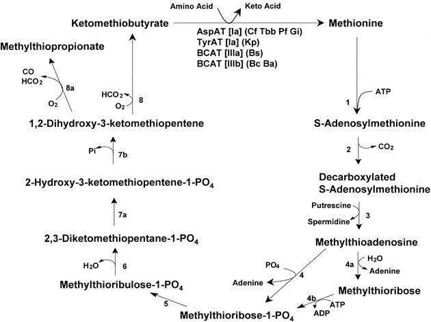
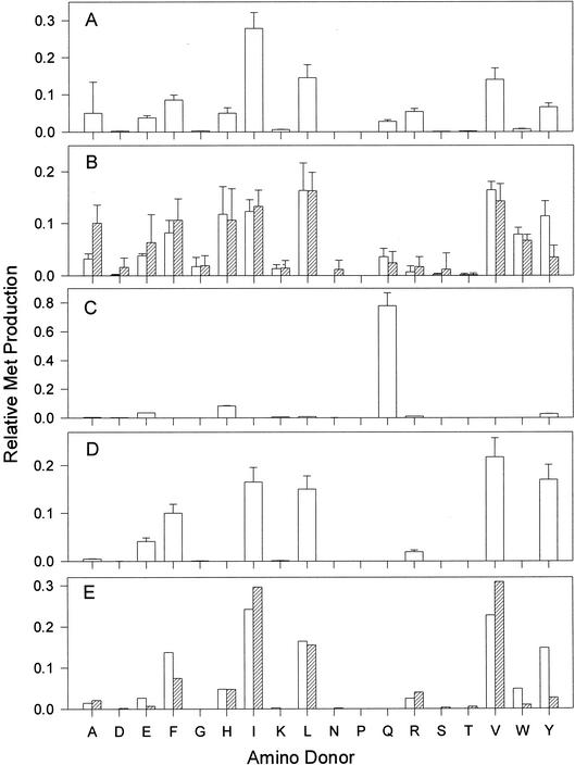
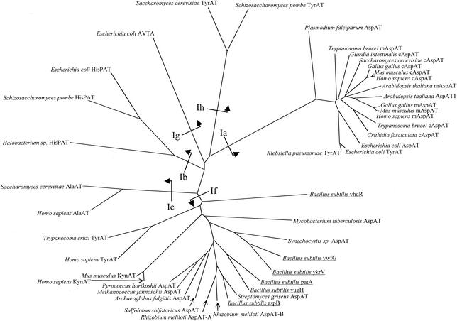
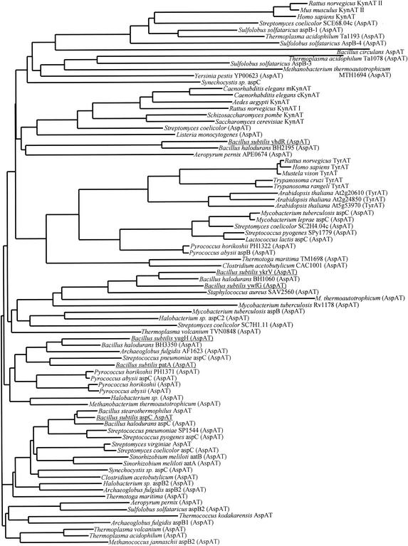
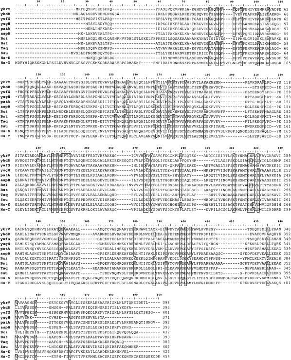
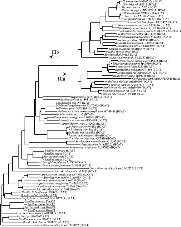
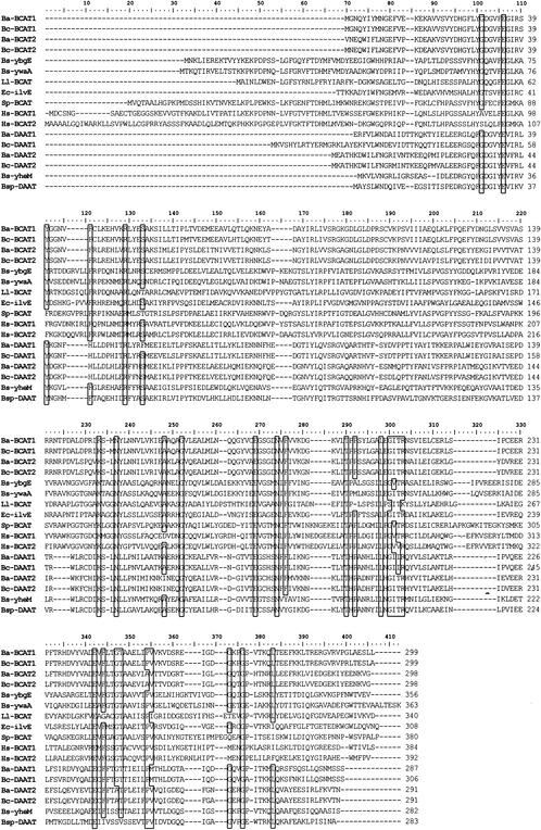
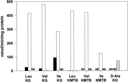
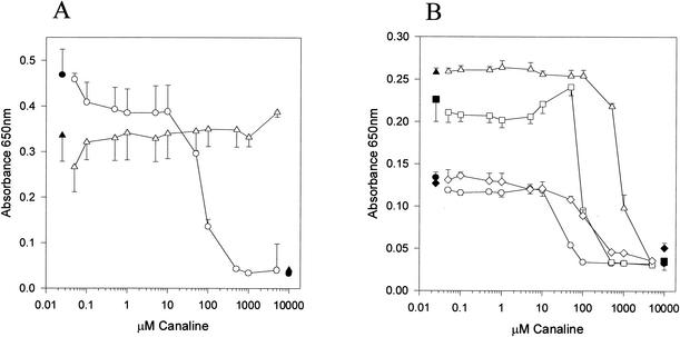
The conversion of ketomethiobutyrate to methionine has been previously examined in a number of organisms, wherein the aminotransferases responsible for the reaction have been found to be members of the Ia subfamily (L. C. Berger, J. Wilson, P. Wood, and B. J. Berger, J. Bacteriol. 183:4421-4434, 2001). The genome of Bacillus subtilis has been found to contain no subfamily Ia aminotransferase sequences. Instead, the analogous enzymes in B. subtilis were found to be members of the If subfamily. These putative aspartate aminotransferases, the yugH, ywfG, ykrV, aspB, and patA gene products, have been cloned, expressed, and characterized for methionine regeneration activity. Only YkrV was able to convert ketomethiobutyrate to methionine, and it catalyzed the reaction only when glutamine was used as amino donor. In contrast, subcellular homogenates of B. subtilis and Bacillus cereus utilized leucine, isoleucine, valine, alanine, phenylalanine, and tyrosine as effective amino donors. The two putative branched-chain aminotransferase genes in B. subtilis, ybgE and ywaA, were also cloned, expressed, and characterized. Both gene products effectively transaminated branched-chain amino acids and ketoglutarate, but only YbgE converted ketomethiobutyrate to methionine. The amino donor preference for methionine regeneration by YbgE was found to be leucine, isoleucine, valine, phenylalanine, and tyrosine. The B. subtilis ybgE gene is a member of the family III of aminotransferases and falls in a subfamily designated here IIIa. Examination of B. cereus and Bacillus anthracis genome data found that there were no subfamily IIIa homologues in these organisms. In both B. cereus and B. anthracis, two putative branched-chain aminotransferases and two putative D-amino acid aminotransferases were discovered as members of subfamily IIIb. These four sequences were cloned from B. cereus, expressed, and characterized. Only the gene product from the sequence designated Bc-BCAT2 was found to convert ketomethiobutyrate to methionine, with an amino donor preference of leucine, isoleucine, valine, phenylalanine, and tyrosine. The B. anthracis homologue of Bc-BCAT2 was also cloned, expressed, and characterized and was found to be identical in activity. The aminooxy compound canaline was found to be an uncompetitive inhibitor of B. subtilis YbgE and also inhibited growth of B. subtilis and B. cereus in culture.


Figures

FIG. 1.
FIG. 1.
The Met regeneration pathway. The labeled enzymes are as follows: 1, S-adenosylmethionine synthetase; 2, S-adenosylmethionine decarboxylase; 3, spermidine/spermine synthetase; 4, methylthioadenosine phosphorylase; 4a, methylthioadenosine nucleosidase; 4b, methylthioribose kinase; 5, unidentified isomerase; 6, unidentified dehydratase; 7, enolase-phosphatase; 8, nonenzymatic or dioxygenase; 8a, dioxygenase. The specific aminotransferases that catalyze the final step are shown, with the subfamily membership in square brackets. The organism abbreviations are as follows: Cf, Crithidia fasciculata; Tbb, Trypanosoma brucei brucei; Pf, Plasmodium falciparum; Gi, Giardia intestinalis; Kp, Klebsiella pneumoniae; Bs, Bacillus subtilis; Bc, Bacillus cereus; Ba, Bacillus anthracis.
FIG. 2.
FIG. 2.
The amino donor range for Met regeneration in Bacillus spp. An enzyme source was mixed with 1.0 mM KMTB, a 2.0 mM concentration of a single amino acid, and pyridoxal phosphate for 30 min at 37°C before analysis of Met production by HPLC. The enzyme sources are as follows: B. subtilis homogenate from cells grown in nutrient broth (A), B. cereus homogenates from cells grown in nutrient broth (open bars) or minimal medium (hatched bars) (B), recombinant B. subtilis YkrV (C), recombinant B. subtilis YbgE (D), or recombinant B. cereus BCAT2 (open bars) or recombinant B. anthracis BCAT2 (hatched bars) (E).
FIG. 3.
FIG. 3.
Family I aminotransferases. Selected sequences were aligned via the clustal algorithm and utilized for tree construction with the neighbor-joining method. The putative B. subtilis AspAT sequences are underlined, and the subfamily designations (as defined by Iraqui et al. [25a]) are shown on the base of the appropriate branches. The enzymes are abbreviated as follows: AspAT, aspartate aminotransferase; TyrAT, tyrosine aminotransferase; AVTA, alanine:valine aminotransferase; HisPAT, histidinol-phosphate aminotransferase; AlaAT, alanine aminotransferase; KynAT, kynurenine aminotransferase.
FIG. 4.
FIG. 4.
The If subfamily of aminotransferases. The sequences were aligned with the clustal algorithm and used for tree construction with the neighbor-joining method. The putative B. subtilis AspATs are underlined.
FIG. 5.
FIG. 5.
Alignment of the putative B. subtilis AspATs. The following sequences were aligned by using the clustal algorithm: B. subtilis YkrV, YhdR, YwfG, YugH, PatA, and AspB, B. circulans (Bci) AspAT (6), Bacillus sp. (Bst) AspAT (49), Thermus aquaticus AspAT (Taq) (38), Sulfolobus solfataricus (Ssu) AspAT (13), human KynAT (Hs-K) (35), and human TyrAT (Hs-T) (47). Residues conserved by 75% of the sequences are boxed.
FIG. 6.
FIG. 6.
Family III aminotransferases. The sequences were aligned with the Clustal algorithm and used for tree construction with the neighbor-joining method. The division between subfamilies IIIa and IIIb is shown by arrows. The B. subtilis, B. cereus, and B. anthracis sequences are underlined.
FIG. 7.
FIG. 7.
Alignment of the B. subtilis, B. cereus, and B. anthracis family III enzymes. The following sequences were aligned with the Clustal algorithm: B. subtilis YbgE, YwaA, and YheM; B. cereus and B. anthracis BCAT1 and -2 and DAAT1 and -2; Ll-BCAT, Lactococcus lactis BCAT (54); Ec-ilvE, E. coli BCAT (29); Sp-BCAT, Schizosaccharomyces pombe BCAT (14); Bsp-DAAT, B. sphaericus DAAT (53); Hs-BCAT1, human BCAT1 (25); Hs-BCAT2, human BCAT2 (15). Residues conserved by 75% of the sequences are boxed.
FIG. 8.
FIG. 8.
Substrate preference for the recombinant B. cereus family III aminotransferases. B. cereus BCAT1 (black bars), BCAT2 (white bars), or DAAT1 (hatched bars) was incubated with 2.0 mM amino acid-1.0 mM keto acid-pyridoxal phosphate before HPLC analysis for the production of Met from KMTB or glutamate from KG. ND, no detectable activity.
FIG. 9.
FIG. 9.
In vitro inhibition of Bacillus sp. growth by canaline. (A) B. subtilis early log cells were inoculated into nutrient broth (triangles) or minimal medium (circles) in the presence of various concentrations of canaline. Growth after incubation overnight at 30°C was measured by turbidity at 650 nm. (B) B. cereus early-log-phase cells were inoculated into nutrient broth (triangles) or minimal medium (circles) in the presence of various amounts of canaline. Growth after incubation overnight at 30°C was measured by turbidity at 650 nm. The squares and diamonds represent minimal medium supplemented with 10 mM Met and 30 mg of BSA/ml, respectively. The dark symbols are the appropriate values for growth with no inhibitor and for medium without cells.

References

    1. Altschul, S. F., T. L. Madden, A. A. Schaffer, J. Zhang, Z. Zhang, W. Miller, and D. J. Lipman. 1997. Gapped BLAST and PSI-BLAST: a new generation of protein database search programs. Nucleic Acids Res. 25:3389-3402. - PMC - PubMed
    1. Anagnostopoulos, C., and J. Spizizen. 1961. Requirements for transformation in Bacillus subtilis. J. Bacteriol. 81:741-746. - PMC - PubMed
    1. Aslanidis, C., and P. J. de Jong. 1990. Ligation-independent cloning of PCR products (LIC-PCR). Nucleic Acids Res. 18:6069-6074. - PMC - PubMed
    1. Bacchi, C. J., J. R. Sufrin, H. C. Nathan, A. J. Spiess, T. Hannan, J. Garofalo, K. Alecia, L. Katz, and N. Yarlett. 1991. 5′-Alkyl-substituted analogs of 5′-methylthioadenosine as trypanocides. Antimicrob. Agents Chemother. 35:1315-1320. - PMC - PubMed
    1. Backlund, P. S., Jr., and R. A. Smith. 1981. Methionine synthesis from 5′-methylthioadenosine in rat liver. J. Biol. Chem. 256:1533-1535. - PubMed

Publication types

MeSH terms

LinkOut - more resources