

Review
doi: 10.1172/JCI18236.
Transplacental thyroxine and fetal brain development
Affiliations
- PMID: 12671044
- PMCID: PMC152596
- DOI: 10.1172/JCI18236
Item in Clipboard
Review
Transplacental thyroxine and fetal brain development
J Clin Invest.
2003 Apr.
No abstract available
Figures
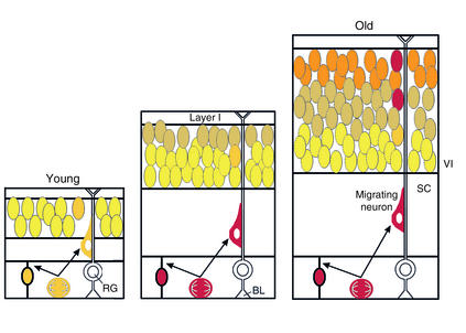
The “inside-out” gradient of cortical lamina is established by neurons migrating farther as development proceeds. Late-born neurons migrate past early-born neurons, forming six layers. Cortical neurons migrate along scaffolds established by radial glia, which are attached to the basal lamina of the ventricular zone and extend through to layer I. Cajal-Retzius neurons in layer I are the first neurons to populate the mantle of the cortex, and they produce and secrete Reelin, which is involved in terminating migration of neurons as they climb the scaffold. The article by Lavado-Autric et al. (1) indicates that thyroid hormone plays a role in the process of neuronal migration. Specifically, rats born to dams with moderately low thyroid hormone have “blurred” cortical lamina, and many neurons do not migrate to their normal destination. Moreover, the action of thyroid hormone in this migration occurs during fetal development. The authors employed this system to show that maternal hypothyroxinemia can produce specific abnormalities in the cytoarchitecture of the cortex during fetal brain development. RG, radial glia; SC, subcortical white matter; BL, basal lamina; VI, layer VI. Adapted with permission from ref. .
Comment on
-
Early maternal hypothyroxinemia alters histogenesis and cerebral cortex cytoarchitecture of the progeny.J Clin Invest. 2003 Apr;111(7):1073-82. doi: 10.1172/JCI16262. J Clin Invest. 2003. PMID: 12671057 Free PMC article.
References
-
- Calvo RM, et al. Fetal tissues are exposed to biologically relevant free thyroxine concentrations during early phases of development. J. Clin. Endocrinol. Metab. 2002;87:1768–1777. - PubMed
-
- Klein, R. 1980. History of congenital hypothyroidism. In Neonatal thyroid screening. G.N. Burrow and J.H. Dussault, editors. Raven Press. New York, New York, USA. 51–59.
-
- Bernal J. Action of thyroid hormone in brain. J. Endocrinol. Invest. 2002;25:268–288. - PubMed
-
- Roti E, Fang S-L, Green K, Emerson CH, Braverman LE. Human placenta is an active site of thyroxine and 3,3′5-triiodothyronine tyrosyl ring deiodination. J. Clin. Endocrinol. Metab. 1981;53:498–501. - PubMed
