. 2003 Apr;111(7):1029-37.
doi: 10.1172/JCI17054.

Rescue of the skeletal phenotype in CasR-deficient mice by transfer onto the Gcm2 null background

Affiliations

Rescue of the skeletal phenotype in CasR-deficient mice by transfer onto the Gcm2 null background

Qisheng Tu et al. J Clin Invest. 2003 Apr.

Abstract

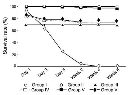
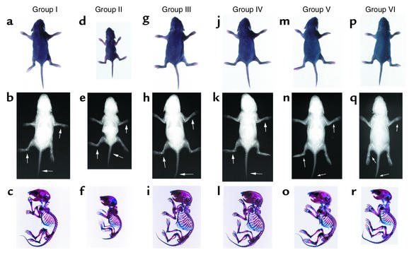
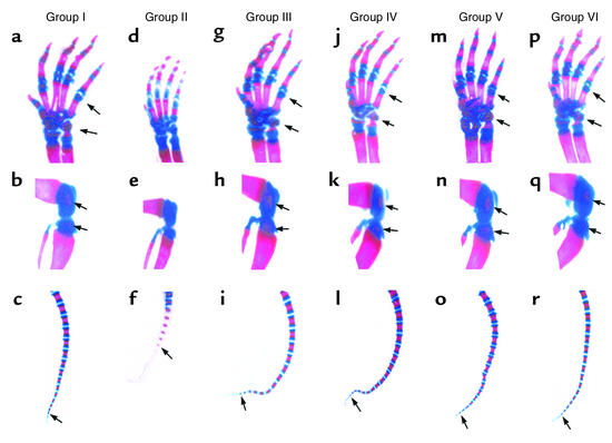
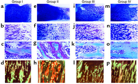
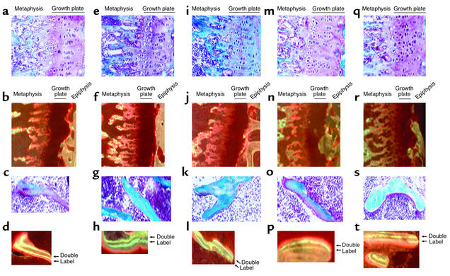
To understand the role of the calcium-sensing receptor (CasR) in the skeleton, we used a genetic approach to ablate parathyroid glands and remove the confounding effects of elevated parathyroid hormone (PTH) in CasR-deficient mice. CasR deficiency was transferred onto the glial cells missing 2-deficient (Gcm2-deficient) background by intercrossing CasR- and Gcm2-deficient mice. Superimposed Gcm2 deficiency rescued the perinatal lethality in CasR-deficient mice in association with ablation of the parathyroid glands and correction of the severe hyperparathyroidism. In addition, the double homozygous CasR- and Gcm2-deficient mice demonstrated healing of the abnormal mineralization of cartilage and bone associated with CasR deficiency, indicating that rickets and osteomalacia in CasR-deficient mice are not due to an independent function of CasR in bone and cartilage but to the effect of severe hyperparathyroidism in the neonate. Analysis of the skeleton of 6-week-old homozygous CasR- and Gcm2-deficient mice also failed to identify any essential, nonredundant role for CasR in regulating chondrogenesis or osteogenesis, but further studies are needed to establish the function of CasR in the skeleton. In contrast, concomitant Gcm2 and CasR deficiency failed to rescue the hypocalciuria in CasR-deficient mice, consistent with direct regulation of urinary calcium excretion by CasR in the kidney. Double Gcm2- and CasR-deficient mice provide an important model for evaluating the extraparathyroid functions of CasR.


Figures

Figure 1
Figure 1
Survival of CasR- and Gcm2-deficient mice. The survival rate of group I, which combines data from heterozygous Gcm2+/– and wild-type mice (n = 38), was 100%, whereas no group II homozygous CasR-deficient mice (n = 29) survived beyond 3 weeks. Group III homozygous Gcm2-deficient mice (n = 13) had a survival rate of 69%. The survival rates of group IV double homozygous CasR- and Gcm2-deficient mice (71%, n = 14) and group VI mixed heterozygous CasR- and homozygous Gcm2-deficient mice (74%, n = 23) are not different from that of group III Gcm2–/– mice. Group V, which combines heterozygous CasR+/– mice and the double heterozygous Gcm2+/–/CasR+/– mice (n = 69), had a survival rate of 97%.
Figure 2
Figure 2
Skeletal phenotype of CasR- and Gcm2-deficient mice. Gross appearance (a, d, g, j, m, and p), skeletal radiographs (b, e, h, k, n, and q), and alizarin red S/Alcian blue–stained whole skeletons (c, f, i, l, o, and r) of 1-week-old mice are shown. The marked reduction in body size, diffuse osteopenia, growth-plate widening (indicated by arrows), and delayed endochondral mineralization in group II homozygous CasR-deficient mice are rescued by superimposed Gcm2 deficiency in group IV double knockout mice. The gross appearance and skeletal phenotype of groups I (normal control), III (homozygous Gcm2-deficient), IV (double homozygous CasR- and Gcm2-deficient), V (double heterozygous CasR- and Gcm2-deficient), and VI (mixed heterozygous CasR- and homozygous Gcm2-deficient mice) are indistinguishable.
Figure 3
Figure 3
Alizarin red S/Alcian blue–stained skeletal regions of CasR- and Gcm2-deficient mice. (ac) Heterozygous Gcm2+/– and wild-type mice. (df) Homozygous CasR-deficient mice. (gi) Homozygous Gcm2-deficient mice. (jl) Double homozygous CasR- and Gcm2-deficient mice. (mo) Heterozygous CasR+/– and double heterozygous Gcm2+/–/CasR+/– mice. (pr) Heterozygous CasR+/– and homozygous Gcm2-deficient mice. Normal appearance of the wrist from a group I control mouse (a) is compared to the widening of the growth plate of the wrist in a 1-week-old group II homozygous CasR-deficient littermate (d). These abnormalities are rescued in group IV double knockout mice (j) and are not present in the other groups (g, m, and p). Mineralization in the epiphysis cartilage of the femur is present in group I control mice (b) but not in the CasR–/– mice (e). Secondary centers of ossification are restored in group IV homozygous CasR- and Gcm2-deficient mice (k). No abnormalities of ossification centers were observed in the other groups (h, n, and q). Endochondral mineralization, as assessed by the number of alizarin red–positive mineralized caudal vertebrae, is reduced in 1-week-old group II CasR–/– mice (f) as compared with group I controls (c), whereas group IV homozygous CasR- and Gcm2-deficient mice (l) as well as the other groups (i, o, and r) show the same number of alizarin red–positive mineralized caudal vertebrae, consistent with a normal temporal and spatial pattern of endochondral bone formation.
Figure 4
Figure 4
Nondecalcified histologic sections of the tibia of 1-week-old CasR- and Gcm2-deficient mice. Shown are group I controls (ad), group II CasR-deficient mice (eh), group III Gcm2-deficient mice (il), and group IV double homozygous CasR- and Gcm2-deficient mice (mp). A toluidine blue–stained section of the growth plate (×125 in a, e, i, and m and ×250 in b, f, j, and n) shows a widened zone of hypertrophic chondrocytes in group II CasR-deficient mice that was corrected in group IV double homozygous CasR- and Gcm2-deficient mice, which were indistinguishable from group I and group III mice. A higher-power view of Goldner-stained sections (×500 in c, g, k, and o) of trabecular bone in secondary spongiosa. Excess osteoid is present in group II CasR–/– mice (g) and resolution of hyperosteoidosis in group IV double knockout mice (o). In Goldner-stained sections, mineralized bone is blue and unmineralized osteoid is reddish-brown in color. The view under fluorescent light of Villanueva-stained sections of metaphysis (×500 in d, h, l, and p) showing the attenuation of calcein deposition in the primary and secondary spongiosa of group II CasR-deficient mice (h) is normalized in group IV double knockout mice (p).
Figure 5
Figure 5
Histologic analysis of the growth plate and bone of 6-week-old homozygous and heterozygous CasR- and Gcm2-deficient mice. Shown are group I normal controls (ad), group III homozygous Gcm2-deficient mice (eh), group IV double homozygous CasR- and Gcm2-deficient mice (il), group V heterozygous Gcm2-deficient mice (mp), and group VI heterozygous CasR- and Gcm2-deficient mice (qt). Goldner-stained sections (×250 in a, e, i, m, and q) show normal growth plates in all groups. Villanueva-stained sections (×125 in b, f, j, n, and r) of the growth plate and adjacent metaphysis show normal mineralization of primary and secondary spongiosa in all groups. Goldner-stained sections (×500 in c, g, k, o, and s) show normal amounts of osteoid in all groups. A highly magnified view (×500 in d, h, l, p, and t) of trabecular bone under fluorescent light reveals normal mineralization in all groups as evidenced by dual calcein and tetracycline labels on the bone surfaces.

Comment in

References

    1. Brown EM, MacLeod RJ. Extracellular calcium sensing and extracellular calcium signaling. Physiol. Rev. 2001;81:239–297. - PubMed
    1. Brown EM, et al. Cloning and characterization of an extracellular Ca2+-sensing receptor from bovine parathyroid. Nature. 1993;366:575–580. - PubMed
    1. Pollak MR, et al. Mutations in the human Ca2+-sensing receptor gene cause familial hypocalciuric hypercalcemia and neonatal severe hyperparathyroidism. Cell. 1993;75:1297–1303. - PubMed
    1. Bai M, et al. Expression and characterization of inactivating and activating mutations in the human Ca2+o-sensing receptor. J. Biol. Chem. 1996;271:19537–19545. - PubMed
    1. Shoback D, Chang W. Starvation amidst plenty — rickets and hypercalcemia in calcium receptor knockout mice. Endocrinology. 2001;142:3733–3735. - PubMed

Publication types

MeSH terms