. 2003 Apr;111(7):1073-82.
doi: 10.1172/JCI16262.

Early maternal hypothyroxinemia alters histogenesis and cerebral cortex cytoarchitecture of the progeny

Affiliations

Early maternal hypothyroxinemia alters histogenesis and cerebral cortex cytoarchitecture of the progeny

Rosalía Lavado-Autric et al. J Clin Invest. 2003 Apr.

Abstract

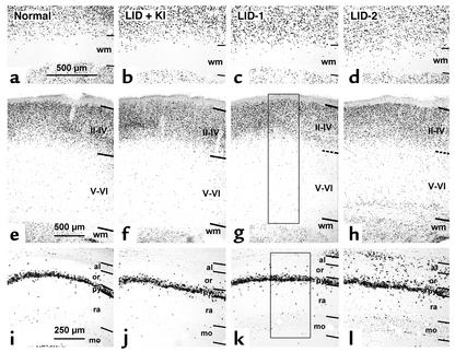
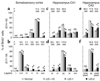
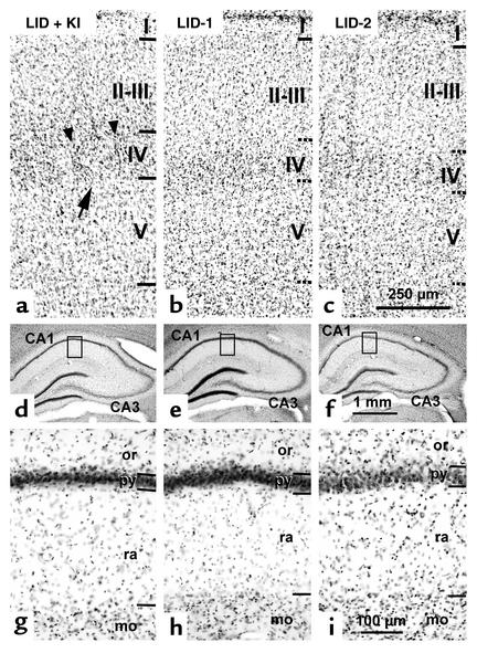
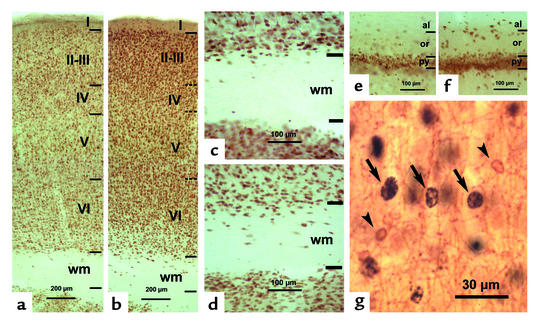
Epidemiological studies from both iodine-sufficient and -deficient human populations strongly suggest that early maternal hypothyroxinemia (i.e., low circulating free thyroxine before onset of fetal thyroid function at midgestation) increases the risk of neurodevelopmental deficits of the fetus, whether or not the mother is clinically hypothyroid. Rat dams on a low iodine intake are hypothyroxinemic without being clinically hypothyroid because, as occurs in pregnant women, their circulating 3,5,3'-triiodothyronine level is usually normal. We studied cell migration and cytoarchitecture in the somatosensory cortex and hippocampus of the 40-day-old progeny of the iodine-deficient dams and found a significant proportion of cells at locations that were aberrant or inappropriate with respect to their birth date. Most of these cells were neurons, as assessed by single- and double-label immunostaining. The cytoarchitecture of the somatosensory cortex and hippocampus was also affected, layering was blurred, and, in the cortex, normal barrels were not formed. We believe that this is the first direct evidence of an alteration in fetal brain histogenesis and cytoarchitecture that could only be related to early maternal hypothyroxinemia. This condition may be 150-200 times more common than congenital hypothyroidism and ought to be prevented both by mass screening of free thyroxine in early pregnancy and by early iodine supplementation to avoid iodine deficiency, however mild.


Figures

Figure 1
Figure 1
Photomicrographs of coronal sections of primary somatosensory cortex (ah) and hippocampus (il) showing BrdU-labeled cells in normal, LID-plus-KI, LID-1, and LID-2 pups at P40. BrdU labeling after E14–E16 (ad) and E17–E19 (el) injections shows that both in the neocortex (ah) and in the hippocampus (il), the radial distribution of BrdU-labeled cells is more widespread in LID-1 and LID-2 pups than in normal and LID-plus-KI pups. Note the increased number of heterotopic BrdU-labeled cells in the white matter of the somatosensory cortex (c and d) and in the strata oriens and alveus of CA1 of the progeny of LID-1 and LID-2 dams (k and l) as compared with the progeny of normal and LID-plus-KI dams (a and b and i and j, respectively). Horizontal lines indicate the borders between layers. Dashed lines indicate borders in g and h, since they are blurred in cresyl violet–stained adjacent sections. Rectangles in g and k show two examples of probes. In each probe, BrdU-labeled cells were plotted, and the relative frequency per layer was calculated. Borders between layers were established by superposing adjacent cresyl violet–stained sections. Strata pyramidale, radiatum, and moleculare are indicated. Magnification for ad is 30×; 17× for eh; and 36× for il. wm, white matter; or, stratum oriens; al, stratum alveus; py, stratum pyramidale; ra, stratum radiatum; mo, stratum moleculare; II–IV, layers II through IV; V–VI, layers V through VI.
Figure 2
Figure 2
Histograms showing the percentage of BrdU-labeled cells in various layers of the primary somatosensory cortex and the CA1 and CA3 regions of the hippocampus. Results for the progeny of the E14–E16 subgroups of normal, LID-plus-KI, LID-1, and LID-2 dams are shown in a, c, and e. Results for the E17–E19 subgroups are shown in b, d, and f. Error bars represent SD across layers from the same group. Single asterisk indicates a significant difference between the LID-1 or LID-2 group and their control group (LID-plus-KI and normal groups); crosshatch indicates a significant difference between the LID-1 group and the LID-2 group. The F and P values corresponding to layers for which significant differences were found between groups are shown in the upper part of each panel. The degrees of freedom were 3, 9 for the E14–E16 subgroups and 3, 7 for the E17–E19 subgroups. inner plexif., inner plexiform stratum; outer plexif., outer plexiform stratum.
Figure 3
Figure 3
Photomicrographs of cresyl violet–stained coronal sections showing the cytoarchitecture of the PMBSF of the primary somatosensory cortex (ac) and hippocampal CA1 (di) in LID-plus-KI, LID-1, and LID-2 progeny at P40. In the PMBSF of LID-plus-KI pups, borders between layers are clear-cut, as expected in normal animals (horizontal lines in a), whereas in LID-1 and LID-2 pups they are blurred (horizontal dashed lines in b and c). In layer IV of LID-plus-KI pups, barrels are normal and well defined, as indicated by an arrow in a, and demarcated by septae (arrowheads). In contrast, barrels in layer IV of LID-1 and LID-2 pups (b and c) are not seen. Enlargements of the insets outlined in d–f are shown in gi. In the hippocampus, the differences between LID-plus-KI pups and both LID-1 and LID-2 pups are not prominent at low magnification (compare d with e and f). However, at a higher magnification, the border between strata pyramidale and oriens in CA1 of LID-plus-KI pups is more clear-cut than in LID-1 and LID-2 progeny (compare g with h and i). Note that strata radiatum is thinner in LID-1 and LID-2 pups than in LID-plus-KI pups. The stratum moleculare is also indicated. Magnification for ac is 54×; 8× for df; and 76× for gi.
Figure 4
Figure 4
Photomicrographs of NeuN-immunostained coronal sections of the primary somatosensory cortex (ad) and hippocampal CA1 (e and f) in LID-plus-KI (a, c, and e) and LID-1 (b, d, and f) progeny at P40. In the neocortex of LID-1 pups, borders between layers are more blurred (horizontal dashed lines in b) than in LID-plus-KI rats (horizontal lines in a). The number of NeuN-labeled neurons increases both in subcortical white matter and in strata oriens and alveus of hippocampal CA1 of LID-1 rats (d and f, respectively) as compared with LID-plus-KI rats (c and e). In g, CNP-positive oligodendrocytes (arrowheads) and BrdU-labeled nuclei (arrows) are shown in layer V of a LID-1 rat from an E14–E16 subgroup. Note that CNP-positive oligodendrocytes are BrdU negative.

Comment in

References

    1. Burrow GN, Fisher DA, Larsen PR. Maternal and fetal thyroid function. N. Engl. J. Med. 1994;331:1072–1078. - PubMed
    1. Morreale de Escobar G, Obregón MJ, Escobar del Rey F. Is neuropsychological development related to maternal hypothyroidism, or to maternal hypothyroxinemia? J. Clin. Endocrinol. Metab. 2000;85:3975–3987. - PubMed
    1. Glinoer D, Delange F. The potential repercussions of maternal, fetal, and neonatal hypothyroxinemia on the progeny. Thyroid. 2000;10:871–877. - PubMed
    1. Calvo RM, et al. Fetal tissues are exposed to biologically relevant free thyroxine concentrations during early phases of development. J. Clin. Endocrinol. Metab. 2002;87:1768–1777. - PubMed
    1. Pop V, et al. Low normal maternal free T4 concentrations during early pregnancy are associated with impaired psychomotor development in infancy. Clin. Endocrinol. (Oxf.) 1999;50:149–155. - PubMed

Publication types

MeSH terms