. 2003 Apr 15;100(8):4545-50.
doi: 10.1073/pnas.0736600100. Epub 2003 Apr 1.

Site-specific contributions to the pH dependence of protein stability

Affiliations

Site-specific contributions to the pH dependence of protein stability

Martin Tollinger et al. Proc Natl Acad Sci U S A. .

Abstract

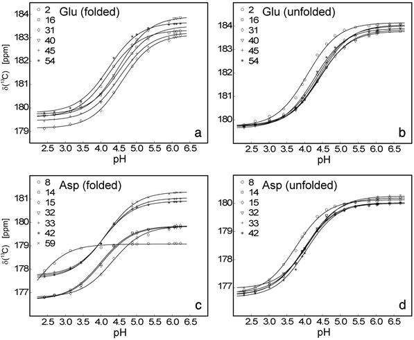
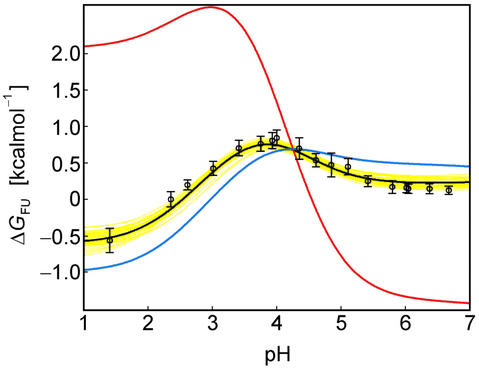
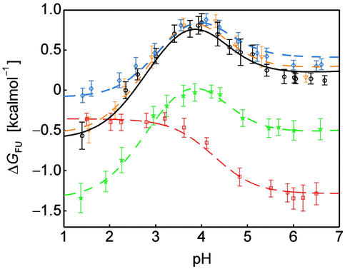
Understanding protein stability is a significant challenge requiring characterization of interactions within both folded and unfolded states. Of these, electrostatic interactions influence ionization equilibria of acidic and basic groups and diversify their pK(a) values. The pH dependence of the thermodynamic stability (Delta G(FU)) of a protein arises as a consequence of differential pK(a) values between folded and unfolded states. Previous attempts to calculate pH-dependent contributions to stability have been limited by the lack of experimental unfolded state pK(a) values. Using recently developed NMR spectroscopic methods, we have determined residue-specific pK(a) values for a thermodynamically unstable Src homology 3 domain in both states, enabling the calculation of the pH dependence of stability based on simple analytical expressions. The calculated pH stability profile obtained agrees very well with experiment, unlike profiles derived from two current models of electrostatic interactions within unfolded states. Most importantly, per-residue contributions to the pH dependence of Delta G(FU) derived from the data provide insights into specific electrostatic interactions in both the folded and unfolded states and their roles in protein stability. These interactions include a hydrogen bond between the Asp-8 side-chain and the Lys-21 backbone amide group in the folded state, which represents a highly conserved interaction in Src homology 3 domains.


Figures

Fig 1.
Fig 1.
Experimental titration curves (carboxyl carbon chemical shifts vs. pH) obtained for glutamate and aspartate side-chain acidic groups in the folded (a and c) and unfolded (b and d) states of the drkN SH3 domain, including best-fit lines (see Methods). Best-fit pKa values are given in Fig. 2c.
Fig 2.
Fig 2.
Analysis of the pH dependence of ΔGFU. (a) Simulation of ΔGformula image(i) for a single ionizable group that titrates with (i) pKformula image = 2.0 and pKformula image = 4.0, orange line, (ii) pKformula image = 4.0 and pKformula image = 2.0, green line, and (iii) pKformula image = pKformula image, dashed black line, at 278 K. (b) The folded state structure of the drkN SH3 domain. Asp and Glu side-chains are color-coded (if |ΔGformula image(i)| > 0.1 kcal⋅mol−1) according to whether pKformula image < pKformula image (orange) or pKformula image < pKformula image (green). (c) Experimental side-chain carboxyl pKa values and error estimates (in parentheses) for Asp and Glu in the Fexch and Uexch states and ΔGformula image(i) values. The pKa values for the side-chain of Asp-59 in the unfolded state (*) could not be obtained due to resonance overlap. (d) Surface electrostatic potential of the drkN SH3 domain at neutral pH, identical view as b. Red represents negative electrostatic potential, white is neutral, and blue represents positive electrostatic potential. (b and d) Diagrams were generated by using the program MOLMOL (38).
Fig 3.
Fig 3.
The pH dependence of protein stability of the drkN SH3 domain between pH 1 and pH 7. Experimentally determined values of ΔGFU (black circles) were determined as ΔGFU = −RT ln(pU/pF), where pU and pF are fractional populations as measured from peak volumes of the Uexch and Fexch states in heteronuclear single quantum coherence experiments and error bars represent SD. The calculated pH dependence of ΔGFU (black line) was derived by using experimental, residue-specific pKa values for both states employing Eqs. 2 and 3. The vertical offset of the calculated curve, ΔGformula image, was determined by minimizing the χ2 with the experimental data. A Monte Carlo simulation was performed by normally distributing pKa values within their SD as estimated from experimental uncertainties (yellow lines). For comparison, pH stability profiles representing simplified models for the unfolded state are plotted assuming (i) standard pKa values (blue line) and (ii) general downward shifted pKa values (by 0.3 pH units from standard values, red line) for the unfolded state but by using experimental pKa values for the folded state.
Fig 4.
Fig 4.
Comparison of the pH stability profiles of the wild-type drkN SH3 domain (black) and mutant proteins His-7 → Ala (blue), Asp-8 → Asn (red), Arg-20 → Ala (green), and Lys-21 → Ala (orange). Values of ΔGFU were determined from peak volumes of the Uexch and Fexch states in heteronuclear single quantum coherence experiments for the wild-type and each mutant, and curves were calculated as described in Methods.

References

    1. Wong K. B., Freund, S. M. & Fersht, A. R. (1996) J. Mol. Biol. 259, 805-818. - PubMed
    1. Mok Y. K., Kay, C. M., Kay, L. E. & Forman-Kay, J. D. (1999) J. Mol. Biol. 289, 619-638. - PubMed
    1. Shortle D. & Ackerman, M. S. (2001) Science 293, 487-489. - PubMed
    1. Klein-Seetharaman J., Oikawa, M., Grimshaw, S. B., Wirmer, J., Duchardt, E., Ueda, T., Imoto, T., Smith, L. J., Dobson, C. M. & Schwalbe, H. (2002) Science 295, 1719-1722. - PubMed
    1. Crowhurst K. A., Tollinger, M. & Forman-Kay, J. D. (2002) J. Mol. Biol. 322, 163-178. - PubMed

Publication types

LinkOut - more resources