. 2003 Apr 15;100(8):4742-7.
doi: 10.1073/pnas.0737855100. Epub 2003 Apr 1.

Treatment with isotretinoin inhibits lipofuscin accumulation in a mouse model of recessive Stargardt's macular degeneration

Affiliations

Treatment with isotretinoin inhibits lipofuscin accumulation in a mouse model of recessive Stargardt's macular degeneration

Roxana A Radu et al. Proc Natl Acad Sci U S A. .

Abstract

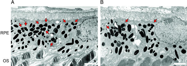
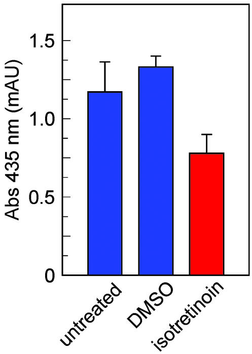
Recessive Stargardt's macular degeneration is an inherited blinding disease of children caused by mutations in the ABCR gene. The primary pathologic defect in Stargardt's disease is accumulation of toxic lipofuscin pigments such as N-retinylidene-N-retinylethanolamine (A2E) in cells of the retinal pigment epithelium. This accumulation appears to be responsible for the photoreceptor death and severe visual loss in Stargardt's patients. Here, we tested a therapeutic strategy to inhibit lipofuscin accumulation in a mouse model of recessive Stargardt's disease. Isotretinoin (Accutane) has been shown to slow the synthesis of 11-cis-retinaldehyde and regeneration of rhodopsin by inhibiting 11-cis-retinol dehydrogenase in the visual cycle. Light activation of rhodopsin results in its release of all-trans-retinaldehyde, which constitutes the first reactant in A2E biosynthesis. Accordingly, we tested the effects of isotretinoin on lipofuscin accumulation in abcr(-/-) knockout mice. Isotretinoin blocked the formation of A2E biochemically and the accumulation of lipofuscin pigments by electron microscopy. We observed no significant visual loss in treated abcr(-/-) mice by electroretinography. Isotretinoin also blocked the slower, age-dependent accumulation of lipofuscin in wild-type mice. These results corroborate the proposed mechanism of A2E biogenesis. Further, they suggest that treatment with isotretinoin may inhibit lipofuscin accumulation and thus delay the onset of visual loss in Stargardt's patients. Finally, the results suggest that isotretinoin may be an effective treatment for other forms of retinal or macular degeneration associated with lipofuscin accumulation.


Figures

Figure 1
Figure 1
Retinoid pathways in the retina and RPE. (A) Visual cycle mediating rhodopsin regeneration. Absorption of a photon (hv) by a rhodopsin molecule in a rod outer-segment disk induces photoisomerization of the 11cRAL chromophore, yielding activated metarhodopsin II. After several seconds, metarhodopsin II decays to yield apo-rhodopsin and free atRAL. Elimination of atRAL from the interior of outer-segment discs is facilitated by the ABCR transporter (11). The atRAL subsequently is reduced to atROL (vitamin A) by atROL dehydrogenase (atRDH). The atROL is released from the outer segment and taken up by an RPE cell, where it is esterified by lecithin retinol acyl transferase (LRAT) to form an all-trans-retinyl ester (atRE). Chemical isomerization is effected by isomerohydrolase (IMH), which uses atRE as a substrate. The resulting 11cROL is oxidized by 11cRDH to form 11cRAL chromophore. 11cRDH is inhibited by isotretinoin with a Ki of ≈0.1 μM (26, 27). The Ki for inhibition of atRDH by isotretinoin is at least two logs higher (N.L.M., R.A.R., and G.H.T., unpublished observations). 11cROL also may serve as a substrate for LRAT to form 11cRE. The final step is recombination of 11cRAL with apo-rhodopsin in the outer segment to form a new molecule of light-sensitive rhodopsin. (B) Synthesis of A2E. After light exposure, newly released atRAL condenses reversibly with phosphatidylethanolamine to form N-ret-PE (step 1). Rarely, a second molecule of atRAL will condense with N-ret-PE to form A2PE-H2 (step 2). The wavelength of maximal absorption (λmax) for A2PE-H2 is 500 nm. Within the acidic and oxidizing environment of RPE phagolysosomes, A2PE-H2 is oxidized to N-retinylidene-N-retinylphosphatidylethanolamine (A2PE; λmax = 435 nm; step 3). Finally, hydrolysis of the phosphate ester yields A2E (λmax = 435 nm) and phosphatidic acid (step 4) (12).
Figure 2
Figure 2
Analysis of visual function by ERG in wild-type and abcr−/− mice after treatment with isotretinoin. (A) b-wave amplitudes elicited in wild-type mice with a dim probe flash (−0.91 log scot td-s) at the indicated times after a 40% photobleach divided by the dark-adapted b-wave amplitude. The mice received a single injection of isotretinoin (40 mg/kg) 1 hr before the photobleach (red circles, n = 7) or received no treatment (blue circles, n = 4). (B) Same protocol as in A except mice were abcr−/− genotype (red circles, n = 4; blue circles, n = 5). (C) RmP3 amplitudes (derived from the leading edge of the a-wave) elicited in wild-type mice with a bright probe flash (3.11 log scot td-s) at the indicated times after a 40% photobleach divided by the dark-adapted RmP3 amplitude. Treatment protocol was similar to A (red circles, n = 7; blue circles, n = 7). (D) Same protocol as in C except mice were abcr−/− genotype (red circles, n = 9; blue circles, n = 9). Error bars show SD.
Figure 3
Figure 3
Effects of isotretinoin on the content of visual retinoids in wild-type and abcr−/− eyes. (A) Levels of 11cRAL in light-adapted, wild-type mice that received either no isotretinoin (blue bar, n = 3 mice) or a single injection 1 hr before tissue collection of isotretinoin at 2.0 (red bar, n = 2), 20 (red bar, n = 3), or 200 (red bar, n = 3) mg/kg. (B) Levels of 11cRE in light-adapted wild-type mice that received either no isotretinoin or a single injection of isotretinoin at the indicated doses (n = 3) as described in A. Note the absence of 11cRE in untreated eyes. (C) [3H]N-ret-PE dpm expressed as a percentage of the total [3H]dpm in phospholipid extracts from eyecups of abcr−/− mice that either were untreated (blue bar, n = 3) or were treated with isotretinoin at 20 mg/kg per day for 10 days (red bar, n = 4). Error bars show SD.
Figure 4
Figure 4
Levels of A2PE-H2 and A2E in abcr−/− RPE after 1 month of treatment with isotretinoin. (A) Levels of A2PE-H2 in RPE from 3-month-old abcr−/− mice before treatment (green bar, n = 3 mice), 4-month-old untreated (blue bar, n = 3) or DMSO-treated (blue bar, n = 3) control mice, and 4-month-old mice treated with 40 mg/kg per day isotretinoin (red bar, n = 3). Values are expressed as peak-height milliabsorption units (mAU) at wavelength (λ) = 500 nm. (B) Levels of A2E in RPE from 3-month-old abcr−/− mice before treatment (green bar, n = 3), 4-month-old DMSO-treated control mice (blue bar, n = 3), and 4-month-old mice treated with 40 mg/kg per day isotretinoin (red bar, n = 3). Values are expressed in mAU at λ = 435 nm. Error bars show SD.
Figure 5
Figure 5
Electron microscopic analysis of RPE from 4-month-old abcr−/− mice. (A) RPE and outer segments (OS) from an untreated mouse. (B) RPE and OS from a mouse treated with isotretinoin for 2 months. Red arrows indicate the irregularly shaped lipofuscin pigment granules, which are distinct from the larger oval melanosomes. (Bars, 2.0 μm.) Note the increased number of lipofuscin granules in RPE from the untreated mouse.
Figure 6
Figure 6
Levels of A2E in 4-month-old wild-type mice after 2 months of treatment with isotretinoin. A2E levels expressed as peak-area mAU (mAU at λ = 435 nm) are shown in RPE from untreated mice (blue bar, n = 4 mice), from mice that received daily injections of DMSO vehicle (blue bar, n = 3), and from mice that received daily injections of isotretinoin at 20 mg/kg (red bar, n = 4). Error bars show SD.

Comment in

References

    1. Leibowitz H M, Krueger D E, Maunder L R, Milton R C, Kini M M, Kahn H A, Nickerson R J, Pool J, Colton T L, Ganley J P, et al. Surv Ophthalmol. 1980;24:335–610. - PubMed
    1. Klein R, Klein B E, Linton K L P. Ophthalmology. 1992;99:933–943. - PubMed
    1. Lee B L, Heckenlively J R. In: Retina-Vitreous-Macula. Guyer D R, Yannuzzi L A, Chang S, Shields J A, Green W R, editors. Philadelphia: Saunders; 1999. pp. 978–988.
    1. Blacharski P A. Fundus flavimaculutus. New York: Raven; 1988.
    1. Eagle R C, Jr, Lucier A C, Bernardino V B, Jr, Yanoff M. Ophthalmology. 1980;87:1189–1200. - PubMed

Publication types

MeSH terms