. 2003 Apr;15(4):899-913.
doi: 10.1105/tpc.009670.

Members of the Arabidopsis dynamin-like gene family, ADL1, are essential for plant cytokinesis and polarized cell growth

Affiliations

Members of the Arabidopsis dynamin-like gene family, ADL1, are essential for plant cytokinesis and polarized cell growth

Byung-Ho Kang et al. Plant Cell. 2003 Apr.

Abstract

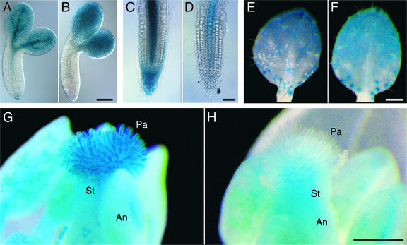
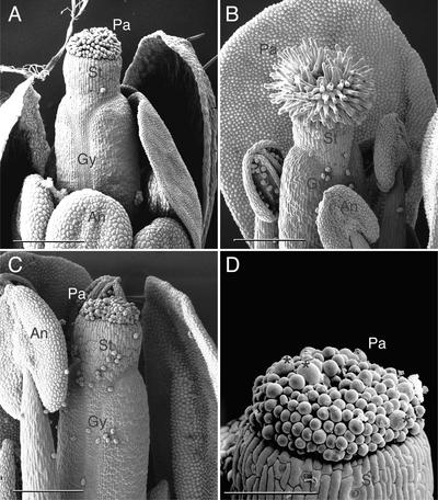
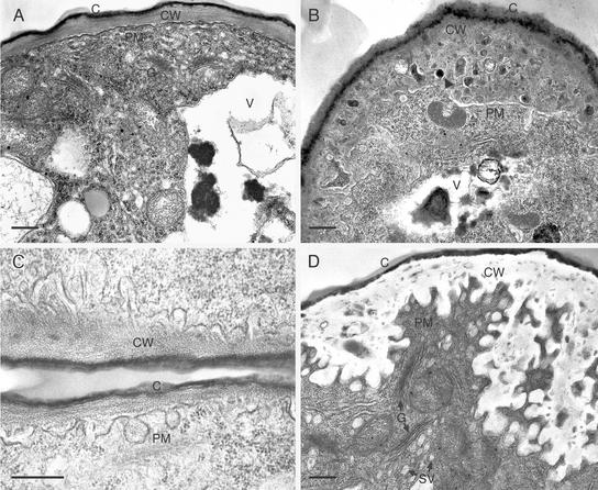
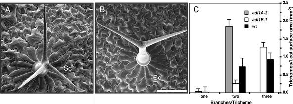
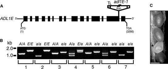
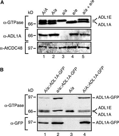
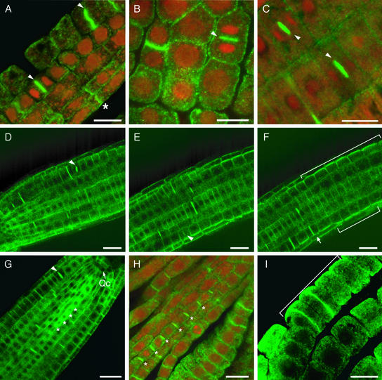
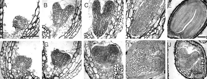
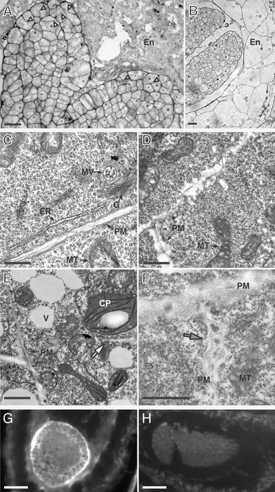
Polarized membrane trafficking during plant cytokinesis and cell expansion are critical for plant morphogenesis, yet very little is known about the molecular mechanisms that guide this process. Dynamin and dynamin-related proteins are large GTP binding proteins that are involved in membrane trafficking. Here, we show that two functionally redundant members of the Arabidopsis dynamin-related protein family, ADL1A and ADL1E, are essential for polar cell expansion and cell plate biogenesis. adl1A-2 adl1E-1 double mutants show defects in cell plate assembly, cell wall formation, and plasma membrane recycling. Using a functional green fluorescent protein fusion protein, we show that the distribution of ADL1A is dynamic and that the protein is localized asymmetrically to the plasma membrane of newly formed and mature root cells. We propose that ADL1-mediated membrane recycling is essential for plasma membrane formation and maintenance in plants.


Figures

Figure 1.
Figure 1.
Histochemical Localization of ADL1A-GUS and ADL1E-GUS Reporter Gene Expression. (A) and (B) Torpedo-stage embryos. (C) and (D) Three-day-old seedling root tips. (E) and (F) Primary leaves of 7-day-old seedlings showing three-branched trichomes. (G) and (H) Mature flowers (stage 14). Processing of ADL1A-GUS ([A], [C], [E], and [G]) and ADL1E-GUS ([B], [D], [F], and [H]) tissue samples for the analysis of GUS activity at each developmental stage was performed in a pair-wise fashion. An, anther; Pa, stigmatic papillae cells; St, stigma. Bars = 100 μm in (B) and (D) and 250 μm in (F) and (H).
Figure 2.
Figure 2.
Scanning Electron Microscopy Analysis of Wild-Type and adl1A-2 Flowers. (A) Dissected stage-12 wild-type flower. Anthers have not dehisced at this stage. (B) Late stage-15 wild-type flower exhibiting dehisced anthers. The gynoecium has begun to elongate rapidly at this stage, presumably as a result of successful self-fertilization. (C) Infertile late stage-16 adl1A-2 flower. The gynoecium has failed to lengthen. (D) Close-up view of adl1A-2 mutant papillae cells showing abnormal isotropically expanded cells (asterisks). An, anther; Gy, gynoecium; Pa, stigmatic papillae cells; St, stigma. Bars = 250 μm in (A) to (C) and 100 μm in (D).
Figure 3.
Figure 3.
Transmission Electron Micrographs of Developing Wild-Type and adl1A-2 Papillae. Longitudinal sections of developing and mature papillae cells are shown. (A) Stigmatic papillae of a wild-type stage-15 flower. (B) and (C) Stigmatic papillae of early stage-11 adl1A-2 flowers. A dark inclusion within the cell wall matrix is indicated by the arrowhead in (B). (D) Stigmatic papillae of a late stage-15 adl1A-2 flower showing PM invaginations. C, cuticle; CW, cell wall; G, Golgi stack; SV, putative secretory vesicle; V, vacuole. Bars = 5 μm in (A) and (B) and 500 nm in (C) and (D).
Figure 4.
Figure 4.
Scanning Electron Microscopy Analysis of Wild-Type and adl1A Leaf Trichomes. (A) and (B) Comparison of trichome branching in wild-type (A) and homozygous adl1A-2 (B) siblings from an adl1A-2/ADL1A heterozygous plant. Sc, socket cell. Bar = 100 μm. (C) Trichome density and the number of branches per trichome were measured on the fourth leaves of wild-type and mutant plants grown under identical conditions. Data are means ± sd of three leaf samples (>125 trichomes per leaf).
Figure 5.
Figure 5.
Homozygous adl1A; E Mutants Display a Synthetic Embryo-Lethal Phenotype. (A) Scheme of the deduced exon/intron structure of ADL1E. Black boxes and lines (drawn to scale) represent exons and introns, respectively. ATG and TGA signify the positions of the translation initiation and termination codons, respectively. The position and orientation of the T-DNA insert (not drawn to scale) in adl1E-1 is shown. Kan, T-DNA neomycin phosphotransferase selectable gene marker; TL, T-DNA left border. (B) Genotypic analysis of the wild type and the adl1A-2, adl1E-1, and adl1A; E mutants. Total DNA was prepared from leaves (lanes 1 to 4) or isolated embryos (lanes 5 to 7) and analyzed by PCR using ADL1A–specific (A), adl1A-2–specific (a), ADL1E–specific (E), and adl1E-1–specific (e) primer sets. PCR products in wild-type [A/A; E/E] (lane 1), adl1A-2 [a/a; E/E] (lane 2), adl1E-1 [A/A; e/e] (lane 3), ADL1A/adl1A-2; ADL1E-1/adl1E-1 [A/a; E/e] (lane 4), ADL1A/ADL1A; adl1E-1/adl1E-1 [A/A; e/e] (lane 5), ADL1A/adl1A-2; adl1E-1/adl1E-1 [A/a; e/e] (lane 6), and adl1A-2/adl1A-2; adl1E-1/adl1E-1 [a/a; e/e] (lane 7) were generated from plants and embryos isolated from self-fertilized heterozygous ADL1A/adl1A-2; ADL1E-1/adl1E-1 plants. (C) Portion of an immature silique (10 to 12 days after flowering) from an ADL1A/adl1A-2; adl1E-1/adl1E-1 plant. Genotypic analysis of genomic DNA prepared from six independent embryos isolated from the pale developing seeds (asterisk) indicated that they were homozygous adl1A; E double mutants, as shown in lane 7 of (B). Bar = 500 μm.
Figure 6.
Figure 6.
Analysis of ADL1A, ADL1E, and ADL1A-mGFP5 Expression in Wild-Type and Mutant Plants. (A) Immunoblot analysis of total protein prepared from rosette leaves of wild-type (lane 1), heterozygous adl1A-2 (lane 2), homozygous adl1A-2 (lane 3), and homozygous adl1E-1 (lane 4) plants. The relative mobilities of the ADL1 proteins in lanes 3 and 4 were confirmed by analysis of a protein mixture containing both homozygous adl1A-2 and homozygous adl1E-1 protein extracts (lane 5). The immunoblots were probed using α-GTPase, α-ADL1A, or α-AtCDC48 antibody. Abbreviations are as in Figure 5. (B) Immunoblot analysis of transgenic lines expressing ADL1A-mGFP5. Total protein was prepared from rosette leaves of transgenic heterozygous adl1A-2 (lane 1), homozygous adl1A-2 (lane 2), and wild-type (lane 4) plants and untransformed heterozygous adl1A-2 plants (lane 3). The immunoblots were probed using α-GTPase or α-GFP antibody.
Figure 7.
Figure 7.
Polarized Localization of ADL1A and ADL1A-mGFP5 in Arabidopsis Roots. (A) Procambial root transition cells stained with affinity-purified α-ADL1A. (B) Lateral root primordium of a homozygous adl1A-2 mutant seedling immunolabeled with affinity-purified GTPase-specific antibodies. (C) ADL1A-mGFP5 in root tissue in chemically fixed homozygous adl1A-2 root cells. (D) to (F) Three frames from the beginning (D), middle (E), and end (F) of the movie (see supplemental data online) showing the localization of ADL1A-mGFP5 in live homozygous adl1A-2 root cells. The arrow in (F) denotes the outer tangential surface of a dividing epidermal cell. (G) ADL1A-mGFP5 localization in the root vascular procambium of a homozygous adl1A-2 seedling. (H) Immunolocalization of ADL1A with α-ADL1A in the root vascular procambium. (I) Immunolocalization of ADL1A with α-ADL1A in the outer tangential surface of epidermal cells in the root elongation zone. ADL1 proteins are shown in green. Nuclei in (A) and (B) are shown in red. (A) to (G) show expanding cell plates in dividing root cells labeled with arrowheads. Asterisks in (A), (G), and (H) indicate ADL1 protein localization in transverse root cell surfaces. Brackets in (F) and (I) indicate the outer tangential surfaces of epidermal cells in the root elongation zone. Qc, quiescent center. Bars = 10 μm in (A), (B), (C), (H), and (I) and 20 μm in (D) to (G).
Figure 8.
Figure 8.
Development of Homozygous adl1A; E Mutant Embryos. Bright-field micrographs of histological sections of morphologically wild-type ([A] to [E]) and abnormal homozygous adl1A; E ([F] to [J]) seeds in the siliques of self-fertilized ADL1A/adl1A; adl1E/adl1E plants at various times during development. Embryos in (A) and (F), (B) and (G), (C) and (H), and (D) and (I) are sibling pairs from the same silique. Developmental stages relative to the wild type (Goldberg et al., 1994) are as follows: (A) and (F), globular; (B) and (G), heart; (C) and (H), torpedo; (D) and (I), walking stick; (E) and (J), mature embryo. Mutant embryos were indistinguishable from wild-type embryos before the globular stage. The arrow in (G) indicates mutant cells that failed to expand anisotropically. Bar in (A) = 50 μm for (A) to (D) and (F) to (I); bar in (E) = 100 μm for (E) and (J).
Figure 9.
Figure 9.
Cellular Morphology of Developing adl1A; E Embryos. (A) and (B) Histological sections of developing mutant (A) and wild-type (B) seeds containing walking stick–stage embryos and endosperm. Arrowheads in (A) indicate cytokinesis-defective protodermal cells with cell wall stubs. (C) to (F) Transmission electron micrographs of phenotypically wild-type (C) and mutant torpedo– (D) and walking stick–stage (E) and (F) embryonic cells. The white arrow in (E) indicates a translucent cell wall stub. The mutant PM is highly irregular and encloses a thickened diffuse cell wall that contains dense inclusions (gray arrow in [F]). (G) and (H) Transverse sections of adl1A; E (G) and phenotypically wild-type (H) sibling embryos stained with the callose binding dye aniline blue. CP, chloroplast; En, endosperm; ER, endoplasmic reticulum; G, Golgi apparatus; MT, mitochondria; MV, multivesicular body; PM, plasma membrane; V, vacuole. Bars = 10 μm in (A) and (B), 200 nm in (B) to (E), and 40 μm in (G) and (H).

References

    1. Arimura, S., and Tsutsumi, N. (2002). A dynamin-like protein (ADL2b), rather than FtsZ, is involved in Arabidopsis mitochondrial division. Proc. Natl. Acad. Sci. USA 99, 5727–5731. - PMC - PubMed
    1. Assaad, F.F., Mayer, U., Wanner, G., and Jürgens, G. (1996). The KEULE gene is involved in cytokinesis in Arabidopsis. Mol. Gen. Genet. 253, 267–277. - PubMed
    1. Battey, N.H., James, N.C., Greenland, A.J., and Brownlee, C. (1999). Exocytosis and endocytosis. Plant Cell 11, 643–660. - PMC - PubMed
    1. Bednarek, S.Y., and Falbel, T.G. (2002). Membrane trafficking during plant cytokinesis. Traffic 3, 621–629. - PubMed
    1. Clough, S.J., and Bent, A.F. (1998). Floral dip: A simplified method for Agrobacterium-mediated transformation. Plant J. 16, 735–743. - PubMed

Publication types

MeSH terms