. 2003 Apr 15;100(8):4474-9.
doi: 10.1073/pnas.0735712100. Epub 2003 Apr 2.

Phosphorylation of p47phox directs phox homology domain from SH3 domain toward phosphoinositides, leading to phagocyte NADPH oxidase activation

Affiliations

Phosphorylation of p47phox directs phox homology domain from SH3 domain toward phosphoinositides, leading to phagocyte NADPH oxidase activation

Tetsuro Ago et al. Proc Natl Acad Sci U S A. .

Abstract

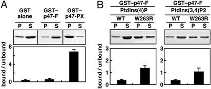
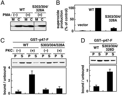
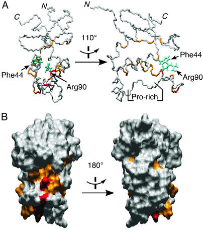
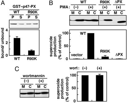
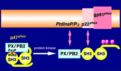
Protein-phosphoinositide interaction participates in targeting proteins to membranes where they function correctly and is often modulated by phosphorylation of lipids. Here we show that protein phosphorylation of p47(phox), a cytoplasmic activator of the microbicidal phagocyte oxidase (phox), elicits interaction of p47(phox) with phosphoinositides. Although the isolated phox homology (PX) domain of p47(phox) can interact directly with phosphoinositides, the lipid-binding activity of this protein is normally suppressed by intramolecular interaction of the PX domain with the C-terminal Src homology 3 (SH3) domain, and hence the wild-type full-length p47(phox) is incapable of binding to the lipids. The W263R substitution in this SH3 domain, abrogating the interaction with the PX domain, leads to a binding of p47(phox) to phosphoinositides. The findings indicate that disruption of the intramolecular interaction renders the PX domain accessible to the lipids. This conformational change is likely induced by phosphorylation of p47(phox), because protein kinase C treatment of the wild-type p47(phox) but not of a mutant protein with the S303304328A substitution culminates in an interaction with phosphoinositides. Furthermore, although the wild-type p47(phox) translocates upon cell stimulation to membranes to activate the oxidase, neither the kinase-insensitive p47(phox) nor lipid-binding-defective proteins, one lacking the PX domain and the other carrying the R90K substitution in this domain, migrates. Thus the protein phosphorylation-driven conformational change of p47(phox) enables its PX domain to bind to phosphoinositides, the interaction of which plays a crucial role in recruitment of p47(phox) from the cytoplasm to membranes and subsequent activation of the phagocyte oxidase.


Figures

Figure 1
Figure 1
(A) Phosphoinositide-binding activity of the full-length p47phox (p47-F). GST–p47-F, GST–p47-PX, or GST alone was incubated with phospholipids/liposomes containing phosphatidylcholine (90%) and PtdIns(4)P (10%). P and S, liposomal pellet and supernatant after centrifugation, respectively. Samples were analyzed by 10% SDS/PAGE, stained with Coomassie brilliant blue, and quantitated by densitometric analysis. The actual bands of GST alone, GST–p47-F, and GST–p47-PX are running at different molecular masses of ≈27, 73, and 40 kDa, respectively. (B) Effect of the W263R substitution on phosphoinositide-binding activity of p47-F. GST–p47-F or GST–p47-F(W263R) was incubated with phospholipids/liposomes containing 10% PtdIns(4)P or PtdIns(3,4)P2. Samples were analyzed as described for A. Each value represents the mean of data from more than three independent experiments, with bars representing the standard deviation.
Figure 2
Figure 2
Phosphorylation-induced conformational change of p47phox, which leads to its interaction with phosphoinositides. (A) Membrane translocation of the wild-type p47phox (WT) and a mutant protein with the S303/304/328A substitution. The gp91phox and p67phox doubly transduced K562 cells were transfected with pREP4 vector or the vector encoding the full-length wild-type p47phox or the one with the S303/304/328A substitution. After cell stimulation with PMA (200 ng/ml), the cell lysates were fractionated by centrifugation, and the cytosolic (C) and membrane (M) fractions were analyzed by immunoblot with anti-p47phox antibody. The actual bands of both wild-type and mutant proteins are running at 47 kDa. (B) Activation of the phagocyte NADPH oxidase by the wild-type p47phox and a mutant protein with the S303/304/328A substitution. Cells were stimulated with PMA as described for A, and superoxide production was determined by superoxide dismutase-inhibitable chemiluminescence change, monitored with DIOGENES. (C) Effect of phosphorylation of p47phox on its phosphoinositide-binding activity. GST–p47-F (WT) or GST–p47-F (S303/304/328A) was treated with PKC in vitro and incubated with phospholipids/liposomes containing 10% PtdIns(4)P. Samples were analyzed as described for Fig. 1B. The actual bands of both GST-fusion proteins are 74 kDa. (D) Effect of the S303/304/328D substitution of p47phox on its phosphoinositide-binding activity. GST–p47-F(WT) or GST–p47-F(S303/304/328D) was incubated with phospholipids/liposomes containing 10% PtdIns(4)P. Samples were analyzed as described for Fig. 1.
Figure 3
Figure 3
NMR analysis of the interaction of the p47phox PX domain and inositol phosphate. The normalized chemical-shift changes were calculated after adding 1.5 mM inositol 1,4,5-trisphosphate to a sample of 0.2 mM 15N-labeled p47phox PX domain. The residues with 1H 15N crosspeaks that shifted >0.065 ppm are shown in orange (Ile-9, Leu-11, Tyr-26, Phe-44, Thr-53, Lys-55, Glu-56, Trp-80, Ala-87, Gln-91, Gly-92, Thr-93, Leu-94, Glu-96, Tyr-97, and His-113), and those that shifted >0.150 ppm (maximum 0.227 ppm) are shown in red (Phe-81, Gly-83, and Arg-90) on the stick drawing (A) and the molecular surface (B) of the p47phox PX structure (PDB ID code 1GD5). The two conserved residues Phe-44 and Arg-90 involved in the inositol phosphate binding are drawn in cyan. (Left) Viewed in the same orientation. (Right) Rotated 110° (A) and 180° (B) along the vertical axis.
Figure 4
Figure 4
Role of the p47phox PX domain and its phosphoinositide-binding activity in activation of the phagocyte NADPH oxidase. (A) Arg-90 of the p47phox PX domain, which is involved in binding to phosphoinositides. GST–p47-PX or GST–p47-PX (R90K) was incubated with phospholipid/liposomes containing 10% PtdIns(3,4)P2. Samples were analyzed as described for Fig. 1. (B) Membrane translocation of the wild-type p47phox (WT), a mutant protein lacking the PX domain (ΔPX), and the one with the R90K substitution and activation of the phagocyte NADPH oxidase in cells expressing these proteins. The gp91phox- and p67phox-transduced K562 cells were transfected with pREP4 vector or the vector to express the indicated form of p47phox. (Upper) After cell stimulation with PMA (200 ng/ml), the cell lysates were fractionated by centrifugation, and the cytosolic fraction (C) and membrane fraction (M) were analyzed by immunoblot with anti-p47phox antibody. The actual bands of p47phox-wild type, p47phox-R90K, and p47phox-ΔPX are running at 47, 47, and 45.5 kDa, respectively. (Lower) Superoxide production was determined by superoxide dismutase-inhibitable chemiluminescence change. (C) Effect of wortmannin on membrane translocation and activation of the oxidase. Cells expressing p47phox-wild type were preincubated in the presence or absence of 100 nM wortmannin (wort) and stimulated with PMA (200 ng/ml). Membrane translocation of p47phox (Left) and superoxide production (Right) were analyzed as described for B.
Figure 5
Figure 5
A proposed mechanism underlying the regulation of p47phox in activation of the phagocyte NADPH oxidase. Human p47phox comprises 390 amino acid residues, and PX/PB2 represents the PX domain (5), also called the PB2 domain (6, 7). The C-terminal SH3 domain, containing Trp-263, interacts intramolecularly with the PXXP motif of the PX domain. Stimulus-induced phosphorylation of p47phox causes a conformational change, by which both PX and SH3 domains become accessible to their membranous targets, phosphoinositides and p22phox, respectively. Cooperation of these two interactions, each being indispensable, enables p47phox to form a stable complex with cytochrome b558 (composed of the two subunit gp91phox and p22phox), leading to activation of the phagocyte NADPH oxidase.

Similar articles

Cited by

References

    1. Fruman D A, Rameh L E, Cantley L C. Cell. 1999;97:817–820. - PubMed
    1. Corvera S, D'Arrigo A, Stenmark H. Curr Opin Cell Biol. 1999;11:460–465. - PubMed
    1. Corvera S, Czech M P. Trends Cell Biol. 1998;8:442–446. - PubMed
    1. Rameh L E, Cantley L C. J Biol Chem. 1999;274:8347–8350. - PubMed
    1. Ponting C P. Protein Sci. 1996;5:2353–2357. - PMC - PubMed

Publication types

MeSH terms

LinkOut - more resources