. 2003 Apr 15;100(8):4445-50.
doi: 10.1073/pnas.0330734100. Epub 2003 Apr 2.

Bioinformatic design of A-kinase anchoring protein-in silico: a potent and selective peptide antagonist of type II protein kinase A anchoring

Affiliations

Bioinformatic design of A-kinase anchoring protein-in silico: a potent and selective peptide antagonist of type II protein kinase A anchoring

Neal M Alto et al. Proc Natl Acad Sci U S A. .

Abstract

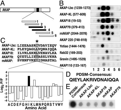
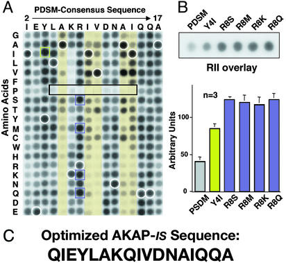
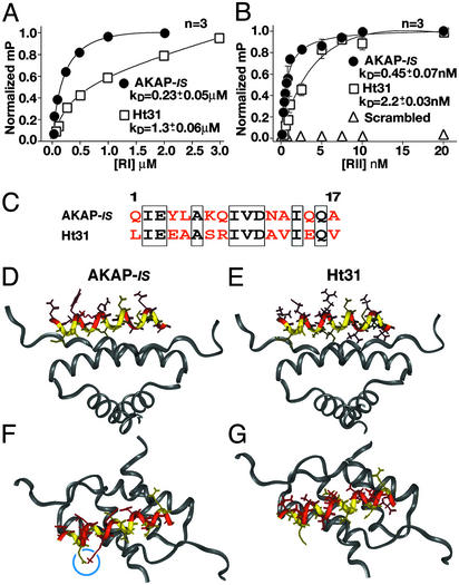
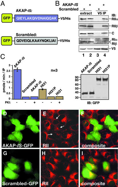
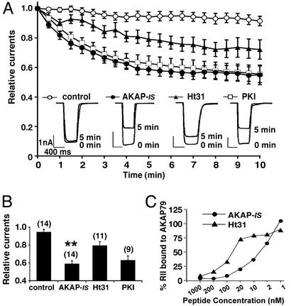
Compartmentalization of the cAMP-dependent protein kinase (PKA) is coordinated through association with A-kinase anchoring proteins (AKAPs). A defining characteristic of most AKAPs is a 14- to 18-aa sequence that binds to the regulatory subunits (RI or RII) of the kinase. Cellular delivery of peptides to these regions disrupts PKA anchoring and has been used to delineate a physiological role for AKAPs in the facilitation of certain cAMP-responsive events. Here, we describe a bioinformatic approach that yields an RII-selective peptide, called AKAP-in silico (AKAP-IS), that binds RII with a K(d) of 0.4 nM and binds RI with a K(d) of 277 nM. AKAP-IS associates with the type II PKA holoenzyme inside cells and displaces the kinase from natural anchoring sites. Electrophysiological recordings indicate that perfusion of AKAP-IS evokes a more rapid and complete attenuation of alpha-amino-3-hydroxy-5-methyl-4-isoxazolepropionic acid (AMPA) receptor currents than previously described anchoring inhibitor peptides. Thus, computer-based and peptide array screening approaches have generated a reagent that binds PKA with higher affinity than previously described AKAPs.


Figures

Figure 1
Figure 1
Derivation of PDSM consensus sequence. (A) Schematic representation of how the RII-binding sequences within AKAPs were defined. Peptide arrays of 20-mer peptides (each offset by three residues) from 10 individual AKAPs were screened for RII binding by the overlay procedure. (B) Autoradiographs of RII-binding peptides from 10 anchoring proteins. The name of each AKAP and segment of sequence analyzed is indicated. (C Upper) Alignment of the five highest-affinity RII-binding sequences by using the MEME software. An AKAP-specific position-dependent scoring matrix (PDSM) was calculated by the log (base 2) of the probability that an amino acid is found at a given position in the alignment divided by the frequency that this amino acid is found in the nonredundant protein database (P/F). (C Lower) Values derived for position 9 in the PDSM sequence represented as a graphical output. Isoleucine (black bar) is the highest-scoring amino acid. Amino acids are indicated by their single-letter codes. (D) The MEME-derived PDSM consensus sequence. (E) RII overlay autoradiograph of a peptide array containing the PDSM consensus sequence and five high-affinity AKAPs (indicated above each lane).
Figure 2
Figure 2
Optimization of PDSM consensus sequence. (A) A two-dimensional array of 320 AKAP-IS peptide derivatives was generated where each residue between positions 2 and 17 in the PDSM sequence (above the array) was replaced with residues having every possible side chain (left of the array). Amino acids are indicated by their single-letter codes. Binding of 32P-labeled RII was detected by autoradiography. Peptide derivatives with substitutions at positions 6, 9, 10, 13, and 14 (yellow columns), proline substitutions with reduced RII binding (black rectangle), and internal control peptides of native sequence (white circles) are indicated. AKAP-IS derivatives with higher apparent RII-binding affinity (green and purple squares) are indicated. (B Upper) Solid-phase RII binding of the original PDSM consensus and the five peptides with higher affinity was quantified by densitometry of autoradiographs. Representative data from three individual experiments are presented. (B Lower) The relative binding affinity (arbitrary units) of each peptide is presented in graphic form. (C) The optimized AKAP-IS sequence.
Figure 3
Figure 3
Biochemical and structural analysis of AKAP-IS. Dissociation constants (Kd) of AKAP-IS (●) and Ht31 (□) peptides were determined by fluorescence polarization. Saturation binding curves were generated with increasing concentrations of RIα (A) or RIIα (B). Polarization values (mP) were determined at equilibrium and normalized to the highest value of saturation. Nonlinear regression analysis was used to derive Kd values from three independent experiments. Interaction of RII was not detected when a scrambled peptide (▵) of amino acid composition identical to that of the AKAP-IS sequence was used. (C) Alignment of the AKAP-IS and Ht31 sequences. Identical (boxed) and dissimilar (red) residues are indicated. (D–G) Molecular modeling of the AKAP-IS–RIIα complex used coordinates from the NMR structure of the Ht31/RIIα complex. The core peptide (yellow) and sites of divergence between AKAP-IS and Ht31 (red) are indicated. A side view reveals a change in RII contact side chains at positions 14 and 17 in AKAP-IS (D) compared with the Ht31 (E). Top view of AKAP-IS (F) reveals the formation of a salt bridge formed by residues E3 and K7 (blue circle) that is not found in Ht31 (G).
Figure 4
Figure 4
AKAP-IS interacts with PKA inside cells. (A) Schematic representation of GFP fusion proteins with the AKAP-IS (Upper) and scrambled (Lower) peptides used in the cell-based studies. Sequences are given by the single-letter amino acid codes. (B) HEK293 cells were transfected with either construct, and immune complexes were immunoprecipitated from cell extracts (extract) with a V5 antibody (V5 IP). Copurification of PKA holoenzyme subunits was detected by immunoblotting (IB) using antibodies against the RIIα (top blot), RIIβ (second blot), C subunit (third blot), or RI subunits (fourth blot). The GFP fusion proteins were detected by immunoblotting using the V5 antibody (bottom blot). (C) The specific activity (pmol/min per IP) of PKA C subunit coprecipitating with chimeric AKAP fusion proteins (indicated above each column) was measured by a filter paper binding assay using the Kemptide as a substrate. PKA activity was blocked when PKI-(5–24) peptide (10 μM) was added to the reaction mixture. The accumulated data from three independent experiments are shown (Left). Immunoblot shows that equal amounts of the GFP fusion proteins were used in these experiments (Right). (D–I) Cells transiently transfected with plasmids expressing AKAP-IS or the scrambled peptide for 24 h were fixed, and immunocytochemical techniques were used to detect intrinsic GFP fluorescence (green; D and G). The subcellular location of RII (red; E and H) was detected with a monoclonal anti-RII antibody and Texas red-conjugated secondary antibody. Arrows indicate the mislocalization of RII from the Golgi/centrosomal area in AKAP-IS-expressing cells. Composite images (F and I) are presented.
Figure 5
Figure 5
AKAP-IS is a potent antagonist of PKA anchoring. (A) Whole-cell patch-clamp recording techniques were used to measure the effects of AKAP-IS (1 μM, ●), Ht31 (1 μM, ▴), or PKI (10 μM, □) peptides on the time-dependent rundown of GluR1 receptor currents expressed in HEK293 cells. The accumulated data from 9–14 experiments including control currents (○) are presented. (Inset) Representative current trace from 0 and 5 min. Amplitude and time scale bars are presented. (B) Graphical representation of the peak current amplitudes upon glutamate stimulation 5 min after delivery of the peptides (indicated below each column). Each bar is normalized to the peak amplitude found at time 0. Amalgamated data from a number of experiments (indicated above each column) are shown. Asterisks indicate significant difference between AKAP-IS and Ht31 (**, P < 0.01). (C) In vitro competition assays. Recombinant AKAP79 (100 ng) immobilized to glutathione-Sepharose was loaded with recombinant 32P-labeled RII. Decreasing concentrations of AKAP-IS (●) or Ht31 (▴) peptide were added. After washing, the remaining RII was detected by autoradiography. RII binding (% bound) and peptide concentrations (nM) are indicated.

References

    1. Sutherland E W. Science. 1972;171:401–408. - PubMed
    1. Lefkowitz R J. J Biol Chem. 1998;273:18677–18680. - PubMed
    1. Taussig R, Gilman A G. J Biol Chem. 1995;270:1–4. - PubMed
    1. Smith F D, Scott J D. Curr Biol. 2002;12:R32–R40. - PubMed
    1. Kaupp U B, Seifert R. Physiol Rev. 2002;82:769–824. - PubMed

Publication types

MeSH terms

Substances

LinkOut - more resources