Review
. 2003 Apr;69(4):1875-83.
doi: 10.1128/AEM.69.4.1875-1883.2003.

Microbiology of the phyllosphere

Affiliations
Review

Microbiology of the phyllosphere

Steven E Lindow et al. Appl Environ Microbiol. 2003 Apr.
No abstract available


Figures

FIG. 1.
FIG. 1.
Fluorescence micrograph of the natural microbial flora colonizing the bean phyllosphere. A large mixed bacterial aggregate (green arrow), which also includes a fungal hypha (orange arrow), has formed at the base of a glandular trichome (white arrow). Bacteria are present also at plant cell junctions and on veins (blue arrows). The red background originates from the autofluorescent chloroplasts within the leaf epidermal cells. The natural microbial flora was visualized by confocal laser scanning microscope-projected z series of the surface of a leaf from a bean plant grown in the field and subsequently incubated under humid conditions at 24°C. The microbial flora was stained with Live BacLight (Molecular Probes, Inc., Eugene, Oreg.), which imparts green and red fluorescence to gram-negative and gram-positive bacteria, respectively. Bar = 20 μm.
FIG. 2.
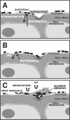
FIG. 2.
Schematic diagram representing various hypothetical bacterial-habitat modifications in the phyllosphere, such as the release of nutrients from plant cells and bacterial cell dispersal effected by the production of syringomycin, which may act both as a phytotoxin and as a surfactant (A); the release of saccharides from the plant cell wall, caused by bacterial secretion of auxin (B); and protection from environmental stresses via production of EPS in bacterial aggregates (C).

References

    1. Andrews, J. H., and R. F. Harris. 2000. The ecology and biogeography of microorganisms on plant surfaces. Annu. Rev. Phytopathol. 38:145-180. - PubMed
    1. Bailey, M. J., P. B. Rainey, X.-X. Zhang, and A. K. Lilley. 2002. Population dynamics, gene transfer and gene expression in plasmids: the role of the horizontal gene pool in local adaptation at the plant surface, p. 173-192. In S. E. Lindow, E. I. Hecht-Poinar, and V. J. Elliot (ed.), Phyllosphere microbiology. APS Press, St. Paul, Minn.
    1. Balandreau, J., V. Viallard, B. Cournoyer, T. Coenye, S. Laevens, and P. Vandamme. 2001. Burkholderia cepacia genomovar III is a common plant-associated bacterium. Appl. Environ. Microbiol. 67:982-985. - PMC - PubMed
    1. Bassler, B. L. 1999. How bacteria talk to each other: regulation of gene expression by quorum sensing. Curr. Opin. Microbiol. 2:582-587. - PubMed
    1. Beattie, G. A. 2002. Leaf surface waxes and the process of leaf colonization by microorganisms, p. 3-26. In S. E. Lindow, E. I. Hecht-Poinar, and V. Elliott (ed.), Phyllosphere microbiology. APS Press, St. Paul, Minn.

LinkOut - more resources