. 2003 Apr;69(4):2230-6.
doi: 10.1128/AEM.69.4.2230-2236.2003.

Surface display of the receptor-binding region of the Lactobacillus brevis S-layer protein in Lactococcus lactis provides nonadhesive lactococci with the ability to adhere to intestinal epithelial cells

Affiliations

Surface display of the receptor-binding region of the Lactobacillus brevis S-layer protein in Lactococcus lactis provides nonadhesive lactococci with the ability to adhere to intestinal epithelial cells

Silja Avall-Jääskeläinen et al. Appl Environ Microbiol. 2003 Apr.

Abstract

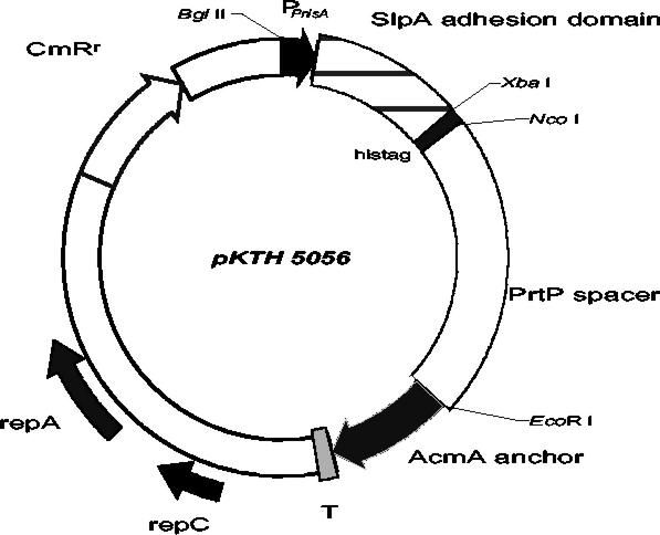
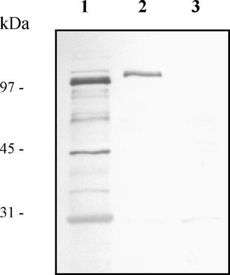
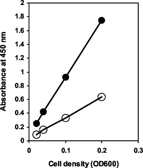
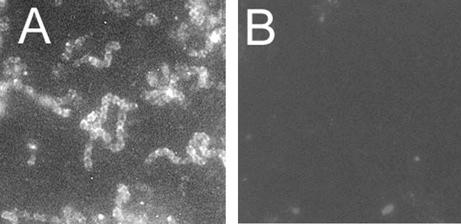
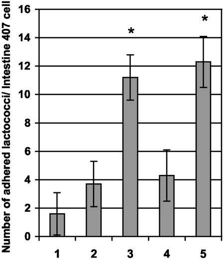
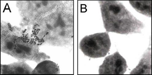
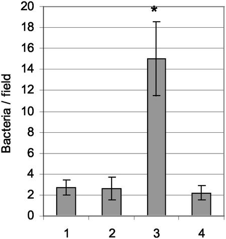
Lactobacillus brevis is a promising lactic acid bacterium for use as a probiotic dietary adjunct and a vaccine vector. The N-terminal region of the S-layer protein (SlpA) of L. brevis ATCC 8287 was recently shown to mediate adhesion to various human cell lines in vitro. In this study, a surface display cassette was constructed on the basis of this SlpA receptor-binding domain, a proteinase spacer, and an autolysin anchor. The cassette was expressed under control of the nisA promoter in Lactococcus lactis NZ9000. Western blot assay of lactococcal cell wall extracts with anti-SlpA antibodies confirmed that the SlpA adhesion domain of the fusion protein was expressed and located within the cell wall layer. Whole-cell enzyme-linked immunosorbent assay and immunofluorescence microscopy verified that the SlpA adhesion-mediating region was accessible on the lactococcal cell surface. In vitro adhesion assays with the human intestinal epithelial cell line Intestine 407 indicated that the recombinant lactococcal cells had gained an ability to adhere to Intestine 407 cells significantly greater than that of wild-type L. lactis NZ9000. Serum inhibition assay further confirmed that adhesion of recombinant lactococci to Intestine 407 cells was indeed mediated by the N terminus-encoding part of the slpA gene. The ability of the receptor-binding region of SlpA to adhere to fibronectin was also confirmed with this lactococcal surface display system. These results show that, with the aid of the receptor-binding region of the L. brevis SlpA protein, the ability to adhere to gut epithelial cells can indeed be transferred to another, nonadhesive, lactic acid bacterium.


Figures

FIG. 1.
FIG. 1.
Schematic drawing of vector pKTH5056, which was constructed for the controlled expression of the surface-anchored adhesion domain of L. brevis SlpA in L. lactis. PnisA, promoter sequence of the nisA gene of L. lactis; AcmA anchor, anchor peptide from the L. lactis acmA gene; PrtP spacer, 515-amino-acid-encoding region of the L. lactis subsp. cremoris Wg2 prtP gene; T, transcription terminator sequence; CmRr, chloramphenicol resistance gene.
FIG. 2.
FIG. 2.
Western blot analysis of the SlpA adhesion-mediating region. Total cellular proteins and cell wall-associated polypeptides were extracted from L. lactis NZ9000 cells harboring pKTH5056. The protein samples were prepared from equal amounts of cells as described in Materials and Methods, and samples were separated by SDS-PAGE, electroblotted onto a nitrocellulose membrane, and incubated with polyclonal SlpA antiserum diluted 1:1,000. Lanes: 1, total cellular proteins from nisin-induced recombinant cells; 2, cell wall-associated polypeptides from nisin-induced recombinant cells; 3, cell wall-associated polypeptides from noninduced recombinant cells. Sizes of molecular mass marker proteins are indicated on the left.
FIG. 3.
FIG. 3.
Whole-cell ELISA for detection of the surface-exposed SlpA adhesion-mediating region encoded by pKTH5056. Anti-SlpA antibody was allowed to bind to lactococcal cells harvested from nisin-induced or uninduced cultures, and after the addition of a horseradish peroxidase conjugate, different lactococcal cell densities were incubated with a chromogenic substrate. Symbols: •, nisin-induced NZ9000(pKTH5056); ○, uninduced NZ9000(pKTH5056). OD600, optical density at 600 nm.
FIG. 4.
FIG. 4.
Immunofluorescence microscopy of recombinant L. lactis NZ9000 cells harboring pKTH5056 (A) and wild-type L. lactis NZ9000 (B). Recombinant cells were harvested after overnight nisin induction, and wild-type cells were harvested after overnight incubation. Both cell types were treated with anti-SlpA antibodies (diluted 1:20 in PBS) and a fluorescein isothiocyanate-conjugated secondary antibody. Both pictures were taken after 16-s exposures. Magnification, ×4,300.
FIG. 5.
FIG. 5.
Adherence of recombinant and wild-type L. lactis NZ9000 cells to Intestine 407 cells. The mean number of adherent lactococcal cells per Intestine 407 cell was determined from 16 randomized microscopic fields and is illustrated for L. lactis NZ9000 cells harboring no plasmid (bar 1), pKTH5046 (nisin induced) (bar 2), pKTH5056 (nisin induced) (bar 3), pKTH5056 (nisin induced and incubated with anti-SlpA antibodies before the adhesion assay) (bar 4), or pKTH5056 (nisin induced and incubated with PBS before the adhesion assay) (bar 5). The results shown are group means with 95% confidence intervals.*, P < 0.01.
FIG. 6.
FIG. 6.
Adhesion of recombinant L. lactis NZ9000 cells carrying pKTH5056 (A) and wild-type L. lactis NZ9000 (B) to Intestine 407 cells. Recombinant lactococcal cells were incubated with PBS for 2 h before the adhesion assay. Magnification, ×4,500.
FIG. 7.
FIG. 7.
Binding of recombinant and wild-type L. lactis NZ9000 to immobilized fibronectin. Means and standard deviations of bacterial numbers in 20 randomly chosen 4 × 102-μm2 microscopic fields are shown for L. lactis NZ9000 harboring no plasmid (bar 1), pKTH5046 (nisin induced) (bar 2), pKTH5056 (nisin induced) (bar 3), or pKTH5056 (uninduced) (bar 4). The concentration of bacteria was 5 × 10⁸ ml−1. The results shown are group means with 95% confidence intervals. *, P < 0.01.

References

    1. Adlerberth, I., S. Ahrné, M-L. Johansson, G. Molin, L. Å. Hanson, and A. E. Wold. 1996. A mannose-specific adherence mechanism in Lactobacillus plantarum conferring binding to the human colonic cell line HT-29. Appl. Environ. Microbiol. 62:2244-2251. - PMC - PubMed
    1. Ausubel, F. M., R. Brent, R. E. Kingston, D. D. Moore, J. G. Seidman, J. A. Smith, and K. Struhl. 1994. Current protocols in molecular biology. Green Publishing Associates, New York, N.Y.
    1. Åvall-Jääskeläinen, S., K. Kylä-Nikkilä, M. Kahala, T. Miikkulainen-Lahti, and A. Palva. 2002. Surface display of foreign epitopes on the Lactobacillus brevis S-layer. Appl. Environ. Microbiol. 68:5943-5951. - PMC - PubMed
    1. Bolken, T. C., C. A. Franke, K. F. Jones, R. H. Bell, R. M. Swanson, D. S. King, V. A. Fischetti, and D. E. Hruby. 2002. Analysis of factors affecting surface expression and immunogenicity of recombinant proteins expressed by gram-positive commensal vectors. Infect. Immun. 70:2487-2491. - PMC - PubMed
    1. Boot, H. J., C. P. A. M. Kolen, J. M. van Noort, and P. H. Pouwels. 1993. S-layer protein of Lactobacillus acidophilus ATCC 4356: purification, expression in Escherichia coli, and nucleotide sequence of the corresponding gene. J. Bacteriol. 175:6089-6096. - PMC - PubMed

Publication types

MeSH terms

LinkOut - more resources