. 2003 Apr;69(4):2313-20.
doi: 10.1128/AEM.69.4.2313-2320.2003.

Heavy metal resistance of biofilm and planktonic Pseudomonas aeruginosa

Affiliations

Heavy metal resistance of biofilm and planktonic Pseudomonas aeruginosa

Gail M Teitzel et al. Appl Environ Microbiol. 2003 Apr.

Abstract

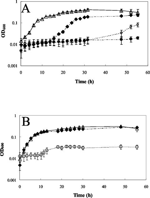
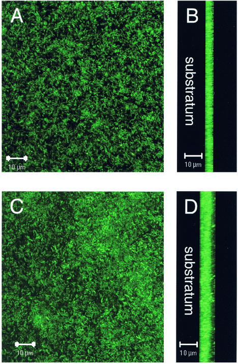
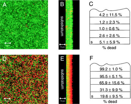
A study was undertaken to examine the effects of the heavy metals copper, lead, and zinc on biofilm and planktonic Pseudomonas aeruginosa. A rotating-disk biofilm reactor was used to generate biofilm and free-swimming cultures to test their relative levels of resistance to heavy metals. It was determined that biofilms were anywhere from 2 to 600 times more resistant to heavy metal stress than free-swimming cells. When planktonic cells at different stages of growth were examined, it was found that logarithmically growing cells were more resistant to copper and lead stress than stationary-phase cells. However, biofilms were observed to be more resistant to heavy metals than either stationary-phase or logarithmically growing planktonic cells. Microscopy was used to evaluate the effect of copper stress on a mature P. aeruginosa biofilm. The exterior of the biofilm was preferentially killed after exposure to elevated concentrations of copper, and the majority of living cells were near the substratum. A potential explanation for this is that the extracellular polymeric substances that encase a biofilm may be responsible for protecting cells from heavy metal stress by binding the heavy metals and retarding their diffusion within the biofilm.


Figures

FIG. 1.
FIG. 1.
Planktonic PAO1 growth in MSVP at 37°C. (A) Growth in the presence of Cu. Symbols: ▵, no added Cu; ⧫, 0.06 mM added Cu; ○, 1 mM added Cu; •, 2 mM added Cu. (B) Growth in the presence of Pb. Symbols: ▵, no added Pb; ⧫, 0.03 mM added Pb; ○, 0.125 mM added Pb.
FIG. 2.
FIG. 2.
Concentrations of free Cu in MSVP and MOPSO-buffered saline. Free Cu concentrations were measured with a Cu-specific electrode. A standard curve was prepared with known Cu concentrations ranging from 0.001 to 10 mM by relating the electrode potential to the concentration of free cationic Cu. Symbols: •, Cu concentration in MOPSO-buffered saline; ○, Cu concentration in MSVP.
FIG. 3.
FIG. 3.
MBCs for biofilm and free-swimming PAO1 cells in response to copper treatment in MOPSO-buffered saline. Biofilm and free-swimming cultures were grown in MSVP at room temperature by using the rotating-disk biofilm reactor. Samples of biofilm and free-swimming cells were harvested and then subjected to a range of Cu concentrations in MOPSO-buffered saline at 37°C for 5 h. Symbols: •, biofilm; ○, free-swimming cells.
FIG. 4.
FIG. 4.
Viability of stationary-phase and logarithmically grown cells subjected to tobramycin stress (A) and copper stress (B). Planktonic cultures were grown at 37°C, and then aliquots were harvested at the logarithmic and stationary phases. Stationary-phase cultures were treated in MSV without a carbon source for 5 h at 37°C, while logarithmic-phase cells were treated in MSVG. In panel A the stationary-phase and logarithmically grown cell points overlap at 2.5 and 3 μg of tobramycin per ml. Symbols: •, logarithmic-phase cells; ○, stationary-phase cells.
FIG. 5.
FIG. 5.
Development of a PAO1 biofilm in MSVP at 30°C. (A and B) Micrographs taken after 2 days of growth. (A) Top down view; (B) saggital view with the substratum to the left. (C and D) Micrographs taken after 5 days of growth. (C) Top down view; (D) saggital view with the substratum to the left. The micrographs were taken with the SCLM by using a magnification of ×630.
FIG. 6.
FIG. 6.
Cu-treated and untreated PAO1 biofilms grown in MSVP at 30°C. (A and B) Micrographs of an untreated biofilm. (A) Top down view; (B) saggital view with the substratum to the left. (D and E) Micrographs of a biofilm treated with 1 mM added Cu for 12 h. (D) Top down view; (E) saggital view with the substratum to the left. Dead cells were stained red with propidium iodide, while live cells were stained green with SYTO9 by using the BacLight LIVE/DEAD viability stain. The micrographs were taken with the SCLM by using a magnification of ×630. (C and F) Quantification of live and dead biomasses as determined by using COMSTAT to estimate the percentage of dead cells as a function of the total biomass within an untreated biofilm (C) and a biofilm treated with 1 mM added copper for 12 h (F). The substratum layer is indicated by s. Averages were calculated for 9 to 12 confocal image stacks for each condition and divided into fifths to normalize for variation in the image stack size. The average depth of the biofilms was 10 μm.

References

    1. Anderl, J. N., M. J. Franklin, and P. S. Stewart. 2000. Role of antibiotic penetration limitation in Klebsiella pneumoniae biofilm resistance to ampicillin and ciprofloxacin. Antimicrob. Agents Chemother. 44:1818-1824. - PMC - PubMed
    1. Bender, C. L., and D. A. Cooksey. 1986. Indigenous plasmids in Pseudomonas syringae pv. tomato: conjugative transfer and role in copper resistance. J. Bacteriol. 165:534-541. - PMC - PubMed
    1. Breault, R. F., J. A. Colman, G. R. Aiken, and D. McKnight. 1996. Copper speciation and binding by organic matter in copper-contaminated streamwater. Environ. Sci. Technol. 30:3477-3486.
    1. Brooun, A., S. Liu, and K. Lewis. 2000. A dose-dependent study of antibiotic resistance in Pseudomonas aeruginosa biofilms. Antimicrob. Agents Chemother. 44:640-646. - PMC - PubMed
    1. Christensen, B. E., J. Kjosbakken, and O. Smidsrød. 1985. Partial chemical and physical characterization of two extracellular polysaccharides produced by marine, periphytic Pseudomonas sp. strain NCMB 2021. Appl. Environ. Microbiol. 50:837-845. - PMC - PubMed

Publication types

MeSH terms

LinkOut - more resources