

A secreted high-affinity inhibitor of human TNF from Tanapox virus
- PMID: 12676996
- PMCID: PMC153641
- DOI: 10.1073/pnas.0737244100
A secreted high-affinity inhibitor of human TNF from Tanapox virus
Abstract
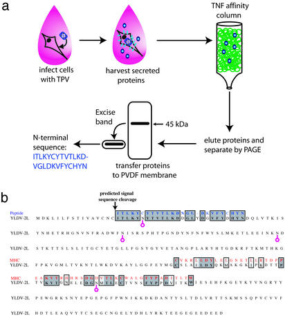
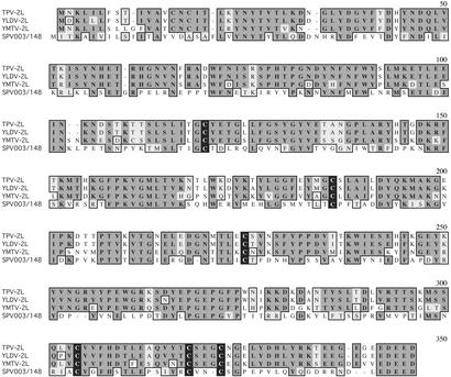
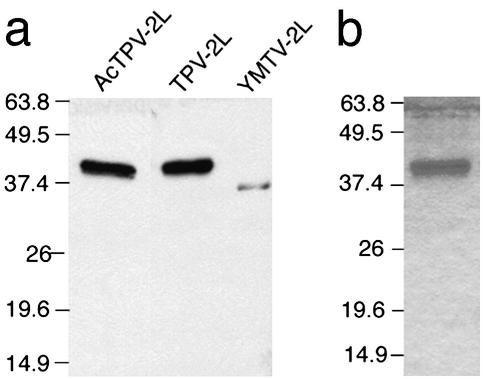
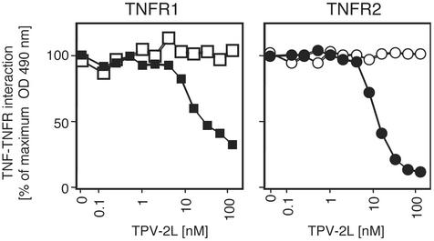
A class of secreted poxvirus tumor necrosis factor (TNF)-binding proteins has been isolated from Tanapox-infected cell supernatants. The inhibitor bound to a TNF-affinity column and was identified as the product of the 2L gene. Sequence analysis of 2L family members from other yatapoxviruses and swinepox virus yielded no sequence homology to any known cellular gene. The expressed Tanapox virus 2L protein bound to human TNF with high affinity (K(d) = 43 pM) and exhibits an unusually slow off-rate. However, 2L is unable to bind to a wide range of human TNF family members. The 2L protein can inhibit human TNF from binding to TNF receptors I and II as well as block TNF-induced cytolysis. Thus, Tanapox virus 2L represents an inhibitor of human TNF and offers a unique strategy with which to modulate TNF activity.
Figures
Similar articles
-
Variation in ligand binding specificities of a novel class of poxvirus-encoded tumor necrosis factor-binding protein.J Biol Chem. 2006 Aug 11;281(32):22517-26. doi: 10.1074/jbc.M604645200. Epub 2006 Jun 16. J Biol Chem. 2006. PMID: 16782702
-
Interaction of human TNF and beta2-microglobulin with Tanapox virus-encoded TNF inhibitor, TPV-2L.Virology. 2009 Apr 10;386(2):462-8. doi: 10.1016/j.virol.2009.01.026. Epub 2009 Feb 20. Virology. 2009. PMID: 19232662
-
Comparative genetic analysis of genomic DNA sequences of two human isolates of Tanapox virus.Virus Res. 2007 Oct;129(1):11-25. doi: 10.1016/j.virusres.2007.05.001. Epub 2007 Jun 14. Virus Res. 2007. PMID: 17574698
-
Tumor necrosis factor inhibitors from poxviruses with an emphasis on tanapoxvirus-2L protein.Recent Pat DNA Gene Seq. 2011 Aug;5(2):97-103. doi: 10.2174/187221511796392033. Recent Pat DNA Gene Seq. 2011. PMID: 21529335 Review.
-
A tumor necrosis factor binding protein (TNF-BP)-physiological antagonist of TNF.Biotherapy. 1991;3(2):159-65. doi: 10.1007/BF02172088. Biotherapy. 1991. PMID: 2054256 Review. No abstract available.
Cited by
-
Tanapoxvirus lacking a neuregulin-like gene regresses human melanoma tumors in nude mice.Virus Genes. 2017 Feb;53(1):52-62. doi: 10.1007/s11262-016-1402-2. Epub 2016 Oct 13. Virus Genes. 2017. PMID: 27738905 Free PMC article.
-
Molluscum contagiosum virus MC80 sabotages MHC-I antigen presentation by targeting tapasin for ER-associated degradation.PLoS Pathog. 2019 Apr 29;15(4):e1007711. doi: 10.1371/journal.ppat.1007711. eCollection 2019 Apr. PLoS Pathog. 2019. PMID: 31034515 Free PMC article.
-
Addition of a Viral Immunomodulatory Domain to Etanercept Generates a Bifunctional Chemokine and TNF Inhibitor.J Clin Med. 2019 Dec 20;9(1):25. doi: 10.3390/jcm9010025. J Clin Med. 2019. PMID: 31877657 Free PMC article.
-
Tanapoxvirus lacking the 15L gene inhibits melanoma cell growth in vitro by inducing interferon-λ1 release.Virus Genes. 2017 Jun;53(3):477-482. doi: 10.1007/s11262-017-1434-2. Epub 2017 Feb 10. Virus Genes. 2017. PMID: 28188458
-
Optimization of codon usage of poxvirus genes allows for improved transient expression in mammalian cells.Virus Genes. 2006 Aug;33(1):15-26. doi: 10.1007/s11262-005-0035-7. Virus Genes. 2006. PMID: 16791414 Free PMC article.
References
-
- Locksley R M, Killeen N, Lenardo M J. Cell. 2001;104:487–501. - PubMed
-
- Beutler B. Tumor Necrosis Factors: The Molecules and Their Emerging Role in Medicine. New York: Raven; 1992.
-
- Chen G, Goeddel D V. Science. 2002;296:1634–1635. - PubMed
-
- Seet B T, Johnston J B, Brunetti C R, Barrett J W, Everett H, Cameron C, Sypula J, Nazarian S, Lucas A, McFadden G. Annu Rev Immunol. 2003;21:377–423. - PubMed
Publication types
MeSH terms
Substances
Associated data
- Actions
- Actions
LinkOut - more resources
Full Text Sources
Other Literature Sources
