. 2003 Apr 15;100(8):4463-8.
doi: 10.1073/pnas.0737647100. Epub 2003 Apr 4.

Amino acids determining enzyme-substrate specificity in prokaryotic and eukaryotic protein kinases

Affiliations

Amino acids determining enzyme-substrate specificity in prokaryotic and eukaryotic protein kinases

Lewyn Li et al. Proc Natl Acad Sci U S A. .

Abstract

The binding between a PK and its target is highly specific, despite the fact that many different PKs exhibit significant sequence and structure homology. There must be, then, specificity-determining residues (SDRs) that enable different PKs to recognize their unique substrate. Here we use and further develop a computational procedure to discover putative SDRs (PSDRs) in protein families, whereby a family of homologous proteins is split into orthologous proteins, which are assumed to have the same specificity, and paralogous proteins, which have different specificities. We reason that PSDRs must be similar among orthologs, whereas they must necessarily be different among paralogs. Our statistical procedure and evolutionary model identifies such residues by discriminating a functional signal from a phylogenetic one. As case studies we investigate the prokaryotic two-component system and the eukaryotic AGC (i.e., cAMP-dependent PK, cGMP-dependent PK, and PKC) PKs. Without using experimental data, we predict PSDRs in prokaryotic and eukaryotic PKs, and suggest precise mutations that may convert the specificity of one PK to another. We compare our predictions with current experimental results and obtain considerable agreement with them. Our analysis unifies much of existing data on PK specificity. Finally, we find PSDRs that are outside the active site. Based on our results, as well as structural and biochemical characterizations of eukaryotic PKs, we propose the testable hypothesis of "specificity via differential activation" as a way for the cell to control kinase specificity.


Figures
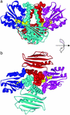
Figure 1
Figure 1
The prokaryotic two-component system and its PSDRs. The x-ray structure (19) of the dimerized catalytic domains (red and cyan), their DDs (red and cyan helices in the center) and the receiver domain of the RR (blue and magenta). The PSDRs are shown as space-filling molecules and are colored the same as the chain in which they occur. A His-30–Asp-54 pair involved in phosphotransfer are shown in the yellow space-filling model. (a) Side view. Note the contacts between PSDRs of the DDs and PSDRs of corresponding RRs (red and magenta, blue and cyan). (b) Top view. Note the contacts between PSDRs of the DDs. Presumably, these interactions are responsible for correct dimerization. Numerous interacting PSDRs also surround the His-30–Asp-54 site.
Figure 2
Figure 2
The eukaryotic PK in the AGC group and its PSDRs. (a) The two-lobe structure of PKA (22) with PSDRs shown by space-fill. The two lobes are red and blue, respectively. The inhibitor substrate (yellow) lies between the lobes. Phospho-Thr-197 is green. (b) PSDRs are as follows: Lys-83, Gln-84, His-87 (blue, upper) Ser 53 (blue, lower) of the PKA and His-23 (yellow) of the inhibitor. Ala-21 (magenta) is the P site. His-23 is the P + 2 site. (c) Phe-129 and Arg-133 (blue) of the PKA, and Arg-18 and Arg-19 on the inhibitor (yellow). The P site is magenta. Arg-18 and Arg-19 correspond to P− 3 and P − 2, respectively. (d) PSDRs Val-191 (red, upper), Tyr-196 (red), Lys-83, and His-87 (blue). Phospho-Thr-197 is green. His-23 and Asp-24 (yellow) are the closest substrate residues to Tyr-196, but are clearly not in contact with Tyr-196.
Figure 3
Figure 3
An illustrative example of how to locate PSDRs from an alignment. The red column has high mutual information and may be a PSDR. The blue and magenta columns have low mutual information and are not PSDRs.
Figure 4
Figure 4
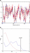
Mutual information (Ii) and its statistical significance. (a) The Ii values for the RR in the prokaryotic two-component system. The abscissa and ordinate represent the sequence position and mutual information, respectively. The blue points are the observed values and the red points are the expected values. The error bars are drawn at three standard deviations from the mean of the expected value and correspond to P = 0.0013. (b) The distribution of Iis for the eukaryotic PKs in the AGC group. The abscissa and ordinate represent, respectively, the mutual information and the probability of observing a particular mutual information. The blue line is the observed result and the red line is the control from the method cited in ref. . The vertical line means that there is a probability of <0.001 of observing an Ii ≥ 1.0 in the control MSAs from the method cited in ref. . Note that “p” in this figure does not correspond to the P value described in the text, which has been determined by the linear transformation method from ref. . See Methods for details.

References

    1. Rubin G M, Yandell M D, Wortman J R, Gabor Miklos G L, Nelson C R, Hariharan I K, Fortini M E, Li P W, Apweiler R, Fleischmann W, et al. Science. 2000;287:2204–2215. - PMC - PubMed
    1. Manning G, Whyte D N, Martinez R, Hunter T, Sudarsanam S. Science. 2002;298:1912–1934. - PubMed
    1. Hunter T. Cell. 1987;50:823–829. - PubMed
    1. Hanks S K, Hunter T. FASEB J. 1995;9:576–596. - PubMed
    1. Maltsev N, Marland E, Yu G-X, Bhatnagar S, Lusk R. Nucleic Acids Res. 2002;30:349–350. - PMC - PubMed

Publication types