

Evidence for a lack of DNA double-strand break repair in human cells exposed to very low x-ray doses
- PMID: 12679524
- PMCID: PMC154297
- DOI: 10.1073/pnas.0830918100
Evidence for a lack of DNA double-strand break repair in human cells exposed to very low x-ray doses
Abstract
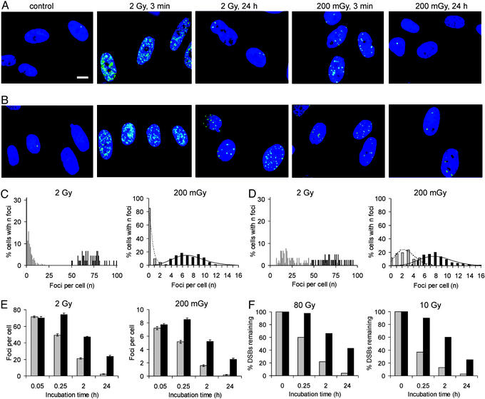
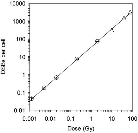
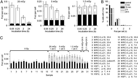
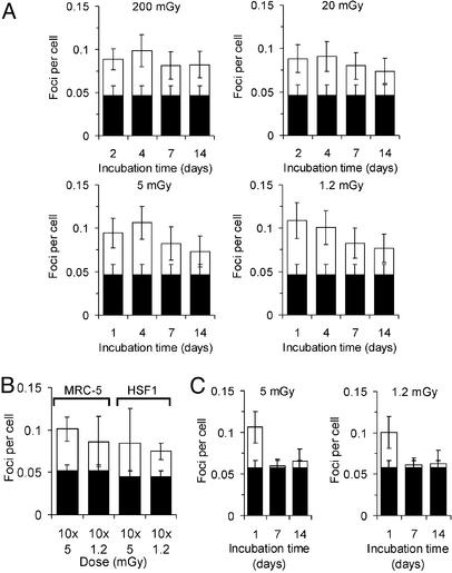
DNA double-strand breaks (DSBs) are generally accepted to be the most biologically significant lesion by which ionizing radiation causes cancer and hereditary disease. However, no information on the induction and processing of DSBs after physiologically relevant radiation doses is available. Many of the methods used to measure DSB repair inadvertently introduce this form of damage as part of the methodology, and hence are limited in their sensitivity. Here we present evidence that foci of gamma-H2AX (a phosphorylated histone), detected by immunofluorescence, are quantitatively the same as DSBs and are capable of quantifying the repair of individual DSBs. This finding allows the investigation of DSB repair after radiation doses as low as 1 mGy, an improvement by several orders of magnitude over current methods. Surprisingly, DSBs induced in cultures of nondividing primary human fibroblasts by very low radiation doses (approximately 1 mGy) remain unrepaired for many days, in strong contrast to efficient DSB repair that is observed at higher doses. However, the level of DSBs in irradiated cultures decreases to that of unirradiated cell cultures if the cells are allowed to proliferate after irradiation, and we present evidence that this effect may be caused by an elimination of the cells carrying unrepaired DSBs. The results presented are in contrast to current models of risk assessment that assume that cellular responses are equally efficient at low and high doses, and provide the opportunity to employ gamma-H2AX foci formation as a direct biomarker for human exposure to low quantities of ionizing radiation.
Figures
Comment in
-
Low-dose radiation: thresholds, bystander effects, and adaptive responses.Proc Natl Acad Sci U S A. 2003 Apr 29;100(9):4973-5. doi: 10.1073/pnas.1031538100. Epub 2003 Apr 18. Proc Natl Acad Sci U S A. 2003. PMID: 12704228 Free PMC article. No abstract available.
References
-
- United Nations Scientific Committee on the Effects of Atomic Radiation. UNSCEAR Sources and Effects of Ionizing Radiation: Report to the General Assembly, with Scientific Annexes. New York: United Nations Scientific Committee on the Effects of Atomic Radiation; 2000.
-
- Hoeijmakers J H J. Nature. 2001;411:366–374. - PubMed
-
- van Gent D C, Hoeijmakers J H J, Kanaar R. Nat Rev Genet. 2001;2:196–206. - PubMed
-
- Rogakou E P, Pilch D R, Orr A H, Ivanova V S, Bonner W M. J Biol Chem. 1998;273:5858–5868. - PubMed
Publication types
MeSH terms
Substances
LinkOut - more resources
Full Text Sources
Other Literature Sources
Medical
