Collagen fiber angle in the submucosa of small intestine and its application in Gastroenterology
- PMID: 12679937
- PMCID: PMC4611454
- DOI: 10.3748/wjg.v9.i4.804
Collagen fiber angle in the submucosa of small intestine and its application in Gastroenterology
Abstract
Aim: To propose a simple and effective method suitable for analyzing the angle and distribution of 2-dimensional collagen fiber in larger sample of small intestine and to investigate the relationship between the angles of collagen fiber and the pressure it undergoes.
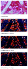
Methods: A kind of 2-dimensional visible quantitative analyzing technique was described. Digital image-processing method was utilized to determine the angle of collagen fiber in parenchyma according to the changes of area analyzed and further to investigate quantitatively the distribution of collagen fiber. A series of intestinal slice's images preprocessed by polarized light were obtained with electron microscope, and they were processed to unify each pixel. The approximate angles between collagen fibers were obtained via analyzing the images and their corresponding polarized light. The relationship between the angles of collagen fiber and the pressure it undergoes were statistically summarized.
Results: The angle of collagen fiber in intestinal tissue was obtained with the quantitative analyzing method of calculating the ratio of different pixels. For the same slice, with polarized light angle's variation, the corresponding ratio of different pixels was also changed; for slices under different pressures, the biggest ratio of collagen fiber area was changed either.
Conclusion: This study suggests that the application of stress on the intestinal tissue will change the angle and content of collagen fiber. The method of calculating ratios of different pixel values to estimate collagen fiber angle was practical and reliable. The quantitative analysis used in the present study allows a larger area of soft tissue to be analyzed with relatively low cost and simple equipment.
Figures


Similar articles
-
Quantitative analysis of collagen fiber angle in the submucosa of small intestine.Comput Biol Med. 2004 Sep;34(6):539-50. doi: 10.1016/j.compbiomed.2003.06.001. Comput Biol Med. 2004. PMID: 15265723
-
The cross-ply arrangement of collagen fibres in the submucosa of the mammalian small intestine.Cell Tissue Res. 1987 Jun;248(3):491-7. doi: 10.1007/BF00216474. Cell Tissue Res. 1987. PMID: 3607846
-
The lattice arrangement of the collagen fibres in the submucosa of the rat small intestine: scanning electron microscopy.Cell Tissue Res. 1988 Jan;251(1):117-21. doi: 10.1007/BF00215455. Cell Tissue Res. 1988. PMID: 3342431
-
Structural and mechanical architecture of the intestinal villi and crypts in the rat intestine: integrative reevaluation from ultrastructural analysis.Anat Embryol (Berl). 2005 Aug;210(1):1-12. doi: 10.1007/s00429-005-0011-y. Epub 2005 Jul 26. Anat Embryol (Berl). 2005. PMID: 16044319
-
[Methods of examining and evaluating morphological changes in small intestine mucosa].Pol Tyg Lek. 1981 Sep 7;36(36):1407-9. Pol Tyg Lek. 1981. PMID: 7036114 Review. Polish. No abstract available.
Cited by
-
Differential biomechanical properties of mouse distal colon and rectum innervated by the splanchnic and pelvic afferents.Am J Physiol Gastrointest Liver Physiol. 2019 Apr 1;316(4):G473-G481. doi: 10.1152/ajpgi.00324.2018. Epub 2019 Jan 31. Am J Physiol Gastrointest Liver Physiol. 2019. PMID: 30702901 Free PMC article.
-
Tissue histology on the correlation between fracture energy and elasticity.Int J Comput Assist Radiol Surg. 2024 Mar;19(3):571-579. doi: 10.1007/s11548-023-03026-6. Epub 2023 Oct 19. Int J Comput Assist Radiol Surg. 2024. PMID: 37855940 Free PMC article.
-
The Macro- and Micro-Mechanics of the Colon and Rectum I: Experimental Evidence.Bioengineering (Basel). 2020 Oct 19;7(4):130. doi: 10.3390/bioengineering7040130. Bioengineering (Basel). 2020. PMID: 33086503 Free PMC article. Review.
-
Morphologic and biomechanical changes of rat oesophagus in experimental diabetes.World J Gastroenterol. 2004 Sep 1;10(17):2519-23. doi: 10.3748/wjg.v10.i17.2519. World J Gastroenterol. 2004. PMID: 15300896 Free PMC article.
-
Visceral pain from colon and rectum: the mechanotransduction and biomechanics.J Neural Transm (Vienna). 2020 Apr;127(4):415-429. doi: 10.1007/s00702-019-02088-8. Epub 2019 Oct 9. J Neural Transm (Vienna). 2020. PMID: 31598778 Free PMC article. Review.
References
-
- Sacks MS, Gloeckner DC. Quantification of the fiber architecture and biaxial mechanical behavior of porcine intestinal submucosa. J Biomed Mater Res. 1999;46:1–10. - PubMed
-
- Clarke KM, Lantz GC, Salisbury SK, Badylak SF, Hiles MC, Voytik SL. Intestine submucosa and polypropylene mesh for abdominal wall repair in dogs. J Surg Res. 1996;60:107–114. - PubMed
-
- Badylak SF, Lantz GC, Coffey A, Geddes LA. Small intestinal submucosa as a large diameter vascular graft in the dog. J Surg Res. 1989;47:74–80. - PubMed
-
- Fackler K, Klein L, Hiltner A. Polarizing light microscopy of intestine and its relationship to mechanical behaviour. J Microsc. 1981;124:305–311. - PubMed
-
- Klein L, Eichelberger H, Mirian M, Hiltner A. Ultrastructural properties of collagen fibrils in rat intestine. Connect Tissue Res. 1983;12:71–78. - PubMed
MeSH terms
Substances
LinkOut - more resources
Full Text Sources

