. 2003 Apr 15;22(8):1725-31.
doi: 10.1093/emboj/cdg183.

Diversification and spectral tuning in marine proteorhodopsins

Affiliations

Diversification and spectral tuning in marine proteorhodopsins

Dikla Man et al. EMBO J. .

Abstract

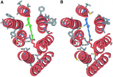
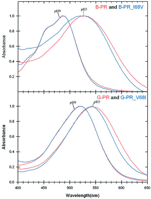
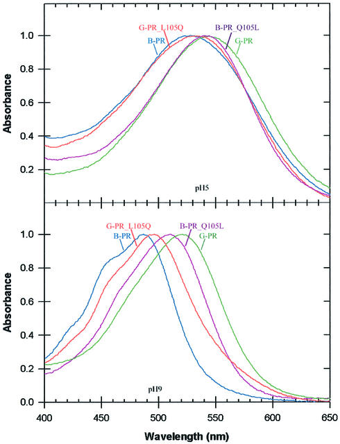
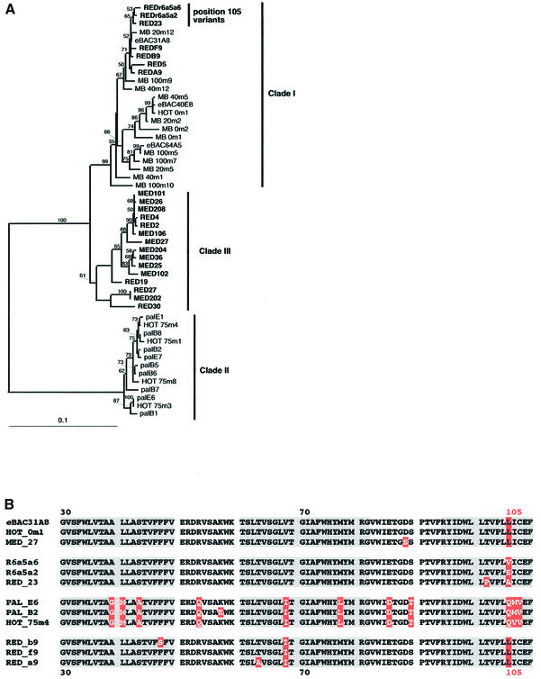
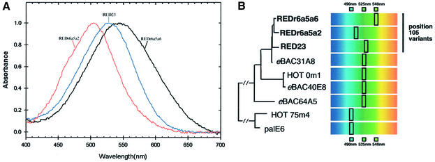
Proteorhodopsins, ubiquitous retinylidene photoactive proton pumps, were recently discovered in the cosmopolitan uncultured SAR86 bacterial group in oceanic surface waters. Two related proteorhodopsin families were found that absorb light with different absorption maxima, 525 nm (green) and 490 nm (blue), and their distribution was shown to be stratified with depth. Using structural modeling comparisons and mutagenesis, we report here on a single amino acid residue at position 105 that functions as a spectral tuning switch and accounts for most of the spectral difference between the two pigment families. Furthermore, looking at natural environments, we found novel proteorhodopsin gene clusters spanning the range of 540-505 nm and containing changes in the same identified key switch residue leading to changes in their absorption maxima. The results suggest a simultaneous diversification of green proteorhodopsin and the new key switch variant pigments. Our observations demonstrate that this single-residue switch mechanism is the major determinant of proteorhodopsin wavelength regulation in natural marine environments.


Figures

None
Fig. 1. Structure modeling of PR (Béjà et al., 2000a) based on the 1.55 Å resolution structure of bacteriorhodopsin (Luecke et al., 1999). (A) Green-absorbing eBAC31A08 variant (G-PR); (B) blue-absorbing PalE6 variant (B-PR). Hydrophilic loops and amino acids in common between the two variants are omitted for clarity. Lys232 in helix G, Schiff base linked to retinal (marked green or blue), is labeled red. Structures were visualized using the ViewerLite 4.2 program (Accelrys Inc.).
None
Fig. 2. Acidic and alkaline absorption spectra of retinal-reconstituted position 68 mutant PRs in E.coli membranes. Spectra were generated by all-trans retinal addition to E.coli membranes containing apoproteins of wild-type and mutant PRs as follows: G-PR, wild-type PR from Monterey Bay (eBAC31A08 in Figure 4; Béjà et al., 2000a); B-PR, wild-type deep-water PR from the HOT station (palE6 in Figure 4; Béjà et al., 2001); BPR_V68I and GPR_V681, V68I and I68V mutants of G-PR and B-PR, respectively. Wild-type PR absorption spectra (in red) and mutant PR spectra (in blue) have been normalized to unity at their absorption maxima for comparison of the spectra.
None
Fig. 3. Acidic and alkaline absorption spectra of retinal-reconstituted position 105 mutant PRs in E.coli membranes. Spectra were generated and normalized as in Figure 2: G-PR, wild-type PR from Monterey Bay (eBAC31A08 in Figure 4; Béjà et al., 2000a); B-PR, deep-water PR from the HOT station (palE6 in Figure 4; Béjà et al., 2001); B-PR_ L105Q and G-PR_Q105L, L105Q and Q105L mutants of G-PR and B-PR, respectively.
None
Fig. 4. Analysis of cloned PR genes from the Red Sea and the Mediterranean Sea. (A) DNA phylogenetic tree based on distance analysis of 740 nt positions by neighbor-joining using the Clustal X (1.81) program (Thompson et al., 1997). Bootstrap values >50% are indicated above the branches. Names in bold type indicate PR variants identified in this study. The scale bar represents number of substitutions per site. All previously reported PR variants have been incorporated in the tree: eBAC31A08–AF279106 (Béjà et al., 2000a) and AF349976–AF350003 (Béjà et al., 2001). (B) Multiple protein alignment of PRs from the different DNA-based groups shown in (A). Residue differences between the protein variants are marked in white on red; position 105 is marked above the alignment and is indicated in all sequences for clarity. Predicted transmembrane helices A, B and C are marked in gray.
None
Fig. 5. pH-dependent absorption spectra of retinal-reconstituted PRs in E.coli membranes. (A) Spectra of deep Red Sea ‘position 105’ variants generated and normalized as in Figure 2 at pH 8. (B) Phylogeny of Red Sea ‘position 105’ PR variants (bold type) and schematic representation of their retinal-reconstituted form λmax in UT5600 E.coli membranes. PRs eBAC31A08, eBAC40E08 and eBAC65A05 were retrieved from surface waters in Monterey Bay (Béjà et al., 2000a, 2001), PR palE6 was collected from Antarctic waters, and HOT 0m1 and HOT 75m4 were collected from Hawaiian waters at 0 and 75 m, respectively (Béjà et al., 2001).

Similar articles

Cited by

References

    1. Béjà O. et al. (2000a) Bacterial rhodopsin: evidence for a new type of phototrophy in the sea. Science, 289, 1902–1906. - PubMed
    1. Béjà O. et al. (2000b) Construction and analysis of bacterial artificial chromosome libraries from a marine microbial assemblage. Environ. Microbiol., 2, 516–529. - PubMed
    1. Béjà O., Spudich,E.N., Spudich,J.L., Leclerc,M. and DeLong,E.F. (2001) Proteorhodopsin phototrophy in the ocean. Nature, 411, 786–789. - PubMed
    1. Béjà O. et al. (2002) Comparative genomic analysis of archaeal genotypic variants in a single population and in two different oceanic provinces. Appl. Environ. Microbiol., 68, 335–345. - PMC - PubMed
    1. Bieszke J.A., Spudich,E.N., Scott,K.L., Borkovich,K.A. and Spudich,J.L. (1999) A eukaryotic protein, NOP-1, binds retinal to form an archaeal rhodopsin-like photochemically reactive pigment. Biochemistry, 38, 14138–14145. - PubMed

Publication types

MeSH terms