. 2003 Apr 15;22(8):1811-23.
doi: 10.1093/emboj/cdg169.

FGFR1 is independently required in both developing mid- and hindbrain for sustained response to isthmic signals

Affiliations

FGFR1 is independently required in both developing mid- and hindbrain for sustained response to isthmic signals

Ras Trokovic et al. EMBO J. .

Abstract

Fibroblast growth factors (FGFs) are signaling molecules of the isthmic organizer, which regulates development of the midbrain and cerebellum. Tissue-specific inactivation of one of the FGF receptor (FGFR) genes, Fgfr1, in the midbrain and rhombomere 1 of the hindbrain of mouse embryos results in deletion of the inferior colliculi in the posterior midbrain and vermis of the cerebellum. Analyses of both midbrain-hindbrain and midbrain-specific Fgfr1 mutants suggest that after establishment of the isthmic organizer, FGFR1 is needed for continued response to the isthmic signals, and that it has direct functions on both sides of the organizer. In addition, FGFR1 appears to modify cell adhesion properties critical for maintaining a coherent organizing center. This may be achieved by regulating expression of specific cell-adhesion molecules at the midbrain-hindbrain border.


Figures

None
Fig. 1. Expression of Fgfr1 and Fgfr2. Whole-mount in situ hybridization analysis of the expression of (AFgfr1 and (BFgfr2 at E7.5. Expression of both genes is detected in the headfolds (arrowheads). In addition, Fgfr1 is strongly expressed in the primitive streak region (PS) and Fgfr2 in the extra-embryonic ectoderm (EE). At E9.5, Fgfr1 is widely expressed (C), whereas Fgfr2 expression is not detected in the anterior rhombomere 1 and the midbrain (D). In situ hybridization analysis of (EFgfr1 and (FFgfr2 expression on sagittal sections of E9.5 embryos. Fgfr1 is widely expressed in the neural tube, whereas Fgfr2 appears to be absent from the tissue around the isthmus. Arrows in (C)–(F) mark the midbrain–hindbrain boundary (MHB).
None
Fig. 2. The conditional Fgfr1 allele, Fgfr1flox, and its inactivation by En1-Cre and Wnt1-Cre. (A) Schematic presentation of the Fgfr1flox allele and its inactivation by the Cre-recombinase. The structures of the FGFR1 protein and the wild-type Fgfr1 locus are shown at the top. Only exons 7, 8, 15 and 16 are depicted. LoxP sites were introduced into introns 7 and 15 by sequential gene targeting to generate the Fgfr1flox allele. Cre-mediated recombination of the Fgfr1flox deletes the transmembrane and most of the intracellular region encoding exons resulting in the inactive Fgfr1Δflox allele. EC, extracellular domain; TM, transmembrane domain; TK, tyrosine kinase domain. To characterize the Cre activity expressed by the En1-Cre and Wnt1-Cre mice, they were crossed with mice carrying a Z/AP reporter allele. (B) Cre-mediated recombination between LoxP sites in the Z/AP allele results in alkaline phosphatase (AP) expression in the midbrain (MB) and rhombomere 1 (R1) of an E8.5 En1-Cre/+; Z/AP/+ embryo. (C) In an E8.5 Wnt1-Cre/+; Z/AP/+ embryo, AP activity is detected in the midbrain and neural crest cells (NCC). Frozen sections of E10.5 (DEn1-Cre/+; Z/AP/+ and (E and FWnt1-Cre/+; Z/AP/+ embryos are shown. In the En1-Cre/+; Z/AP/+ embryos, AP was expressed in the midbrain and rhombomere 1. In Wnt1-Cre/+; Z/AP/+ embryos, AP activity was detected in the midbrain and scattered cells of the rhombomere 1. A boundary is observed between the AP-positive midbrain and mostly AP-negative rhombomere 1 (F). Arrowheads in (B)–(F) point to the midbrain–hindbrain boundary. (GL) Inactivation of Fgfr1 expression by En1-Cre and Wnt1-Cre. Whole-mount in situ hybridization analysis of E8.5 (10 somite stage) En1-Cre/+; Fgfr1flox/flox (G) and wild-type (WT) embryos reveals inactivation of Fgfr1 transcription in the midbrain–hindbrain region by En1-Cre (arrowheads). In situ hybridization analysis of Fgfr1 expression in sagittal sections of E9.5 En1-Cre/+; Fgfr1flox/flox (H), Wnt1-Cre/+; Fgfr1flox/Δflox (I) and wild-type (K) embryos. In En1-Cre/+; Fgfr1flox/flox embryos, inactivation of Fgfr1 expression occurs both in the midbrain and rhombomere 1, whereas in Wnt1-Cre/+; Fgfr1flox/Δflox embryos rhombomere 1 still expresses Fgfr1. Regions of affected Fgfr1 expression are indicated by arrowheads (H and I). A parallel section to the one shown in (I) hybridized with the Fgf8 probe (L). A double-headed arrow in (H)–(L) marks the midbrain–hindbrain boundary.
None
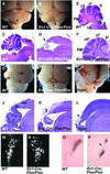
Fig. 3. Morphology of the midbrain and cerebellum of the midbrain–hindbrain-specific and hypomorphic Fgfr1 mutants. Whole-mount view of (A) wild-type and (BEn1-Cre/+; Fgfr1flox/flox adult brains. The entire vermis is missing in En1-Cre/+; Fgfr1flox/flox mice (arrow). (C and D) Midsagittal and (E and F) parasagittal sections of adult wild-type (C and E) and En1-Cre/+; Fgfr1flox/flox (D and F) brains. Complete aplasia of the vermis is evident in the En1-Cre/+; Fgfr1flox/flox mice [arrow in (D)]. The En1-Cre/+; Fgfr1flox/flox mice have cerebellar hemispheres, but their pattern of foliation is altered (F). Whole-mount views and corresponding midsagittal sections of new-born wild-type (G and J), En1-Cre/+; Fgfr1flox/flox (H and K) and Fgfr1n15YF/n15YF (I and L) mice. Aplasia of the vermis and deletions of the inferior colliculi can be seen in both types of Fgfr1 mutants (arrows). Locus coeruleus, identified by Dopamine-β-hydroxylase mRNA in situ hybridization of adult brain sections (M and N) and anti-tyrosine hydroxylase immuno staining of newborn brain sections (O and P) appears disorganized in the En1-Cre/+; Fgfr1flox/flox mutants (N and P) compared with wild type (M and O). cI, crus I; cII, crusII; ch, cerebellar hemisphere; cp, choroids plexus; ic, inferior colliculus; mb, midbrain; PM, paramedian lobule; S, simplex; v, vermis. The mutant hemisphere lobes are labeled with asterisks.
None
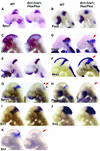
Fig. 4. Analysis of gene expression in the midbrain–hindbrain specific Fgfr1 mutants. Whole-mount in situ hybridization of E8.5 (A and B), E9.5 (CJ) and E10 (K) wild-type and En1-Cre/+; Fgfr1flox/flox embryos with Sprouty1 (A, G), Pax2 (B, H), Otx2 (C), Gbx2 (D), Fgf8 (E), Wnt1 (F), En1 (I, K) and En2 (J) probes. Red arrows indicate altered gene expression. Small arrowheads in (G) indicate remaining Sprouty1 expression in regions distal to the isthmus. See text for details.
None
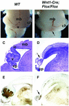
Fig. 5. Morphology of the midbrain and cerebellum of the midbrain-specific Fgfr1 mutants. Whole-mount views (A and B) and mid-sagittal sections (C and D) of brains of newborn wild-type (A and C) and Wnt1-Cre/+; Fgfr1flox/flox (B and D) mice. Calbindin expression in wild-type (E) and Wnt1-Cre/+; Fgfr1flox/flox mice (F). Arrows indicate the deletion of the inferior colliculi and malformed vermis. ch, cerebellar hemisphere; cp, choroid plexus; mb, midbrain; v, vermis.
None
Fig. 6. Analysis of gene expression in the midbrain specific Fgfr1 mutants. Whole-mount in situ hybridization of E9.5 (A–J) and E10.0 (K) wild-type and Wnt1-Cre/+; Fgfr1flox/Δflox embryos with Otx2 (A and B), Fgf8 (C and D), Wnt1 (E and F), Sprouty1 (G), Pax2 (H), En1 (I and K) and En2 (J) probes. Close-up side views of embryos hybridized with Otx2 (B) and Wnt1 (F), as well as a slightly oblique dorsal view of embryos hybridized with Fgf8 probe (D). Arrowheads indicate the isthmus, red arrows indicate altered gene expression. See text for details.
None
Fig. 7. Expression of Ephrin-A5 and PB-cadherin in Fgfr1 mutants and a model for FGFR1 function during the maintenance of the isthmic organizer. Whole-mount in situ hybridization analysis of Ephrin-A5 (A, B) and PB-cadherin (CE) expression in E9.5 wild-type (A, C), En1-Cre/+; Fgfr1flox/flox (B, D) and Wnt1-Cre/+; Fgfr1flox/Δflox (E) embryos. In both types of Fgfr1 mutants PB-cadherin expression is down-regulated in the midbrain, especially in its dorsal part. Arrowheads in (A)–(E) point to the midbrain–hindbrain boundary. (F) A model for the function of FGF signaling through FGFR1 during maintenance of the isthmic organizer. We suggest that FGFR1 regulates gene expression independently in the midbrain (Mes) and hindbrain (Met). Some of the FGFR1-regulated genes confer specific adhesive characteristics to the cells next to the midbrain–hindbrain border.

Similar articles

Cited by

References

    1. Acampora D., Avantaggiato,V., Tuorto,F. and Simeone,A. (1997) Genetic control of brain morphogenesis through Otx gene dosage requirement. Development, 124, 3639–3650. - PubMed
    1. Ang S.L. and Rossant,J. (1993) Anterior mesendoderm induces mouse Engrailed genes in explant cultures. Development, 118, 139–149. - PubMed
    1. Bally-Cuif L., Cholley,B. and Wassef,M. (1995) Involvement of Wnt-1 in the formation of the mes/metencephalic boundary. Mech. Dev., 53, 23–34. - PubMed
    1. Blair S.S. and Ralston,A. (1997) Smoothened-mediated Hedgehog signalling is required for the maintenance of the anterior–posterior lineage restriction in the developing wing of Drosophila. Development, 124, 4053–4063. - PubMed
    1. Broccoli V., Boncinelli,E. and Wurst,W. (1999) The caudal limit of Otx2 expression positions the isthmic organizer. Nature, 401, 164–168. - PubMed

Publication types

MeSH terms