. 2003 Apr 15;22(8):1846-56.
doi: 10.1093/emboj/cdg179.

Chromatin remodeling protein Chd1 interacts with transcription elongation factors and localizes to transcribed genes

Affiliations

Chromatin remodeling protein Chd1 interacts with transcription elongation factors and localizes to transcribed genes

Rajna Simic et al. EMBO J. .

Abstract

Transcription in eukaryotes is influenced by the chromatin state of the template, and chromatin remodeling factors have well-documented roles in regulating transcription initiation by RNA polymerase (pol) II. Chromatin also influences transcription elongation; however, little is known about the role of chromatin remodeling factors in this process. Here, we present evidence that the Saccharomyces cerevisiae chromatin remodeling factor Chd1 functions during transcription elongation. First, we identified Chd1 in a two-hybrid screen for proteins that interact with Rtf1, a member of the Paf1 complex that associates with RNA pol II and regulates transcription elongation. Secondly, we show through co-immunoprecipitation studies that Chd1 also interacts with components of two essential elongation factors, Spt4-Spt5 and Spt16-Pob3. Thirdly, we demonstrate that deletion of CHD1 suppresses a cold-sensitive spt5 mutation that is also suppressed by defects in the Paf1 complex and RNA pol II. Finally, we demonstrate that Chd1, Rtf1 and Spt5 associate with actively transcribed regions of chromatin. Collectively, these findings suggest an important role for Chd1 and chromatin remodeling in the control of transcription elongation.


Figures

None
Fig. 1. Identification of Chd1 as an Rtf1-interacting protein. (A) Diagram of the wild-type Chd1 protein and deletion derivatives used in the analysis shown in Table I and Figure 2. The plasmids encoding these different forms of Chd1 are: CHD1, pGH269; ΔSnaBI, pGH272; ΔStuI, pGH273; ΔSphI, pGH274; and ΔChr, pGH271. The Gal4–Chd1 fusion protein identified in the two-hybrid screen with Rtf1 contains amino acids 863 (arrow) to 1468. (B) β-galactosidase assays were performed on transformants of yeast strain PJ69-4A that expressed the indicated Gal4 fusion proteins (GBD, Gal4-DNA binding domain; GAD, Gal4 activation domain). The following plasmids were used: pLS28 (GBD-Rtf1), pKA202 (original library isolate of GAD-Chd1), pGBT9 and pGAD424. For each plasmid combination, the reported units are mean values from assays performed on three independent transformants at two different extract concentrations. Error bars denote standard errors. (C) Chd1 co-immunoprecipitates with Rtf1. Anti-Rtf1 or pre-immune serum was added to extracts prepared from yeast strain GHY773 for co-immunoprecipitation analysis. Precipitated proteins were analyzed by immunoblotting with anti-Rtf1 and anti-HA1 antisera. Lane 1, 20 µg of whole-cell extract; lanes 2 and 4, 20 µg of unbound material; lanes 3 and 5, total bound material.
None
Fig. 2. Spt5 and Chd1 interactions. (Achd1Δ mutations suppress an spt5 Cs mutation. A chd1Δ spt5 Cs strain (GHY305) was transformed with plasmids encoding wild-type Chd1 (pGH269), Chd1 with the K407R substitution (pGH268), a derivative of Chd1 lacking both chromodomains (pGH271), and an empty URA3 vector (pRS316). Five-fold dilutions of the transformants were plated to two SC-uracil plates. One of the plates was incubated at 15°C for 6 days and the other was incubated at 30°C for 2 days. (B) Chd1 co-immunoprecipitates with Spt5-Flag. Anti-Flag immunoprecipitations were performed on extracts of an Spt5-Flag strain (GHY617) and an untagged control strain (mock, GHY611). Precipitated proteins were analyzed by immunoblotting with anti-Chd1 antisera (Tran et al., 2000). Lanes: WCE, 30 µg of the Spt5-Flag extract; Mock, the untagged immunoprecipitate; Spt5-Flag, the Spt5-Flag immunoprecipitate. (C) Spt5 and Pob3 co-immunoprecipitate with HA3-Chd1. Extracts of a HA3-Chd1strain (GHY773) and an untagged control strain (mock; FY119) were immunoprecipitated with anti-HA antisera. Precipitated proteins were analyzed by immunoblotting with anti-Spt5 and anti-Pob3 antisera.
None
Fig. 3. Rtf1 and Spt5 associate with the coding regions of genes. (A) ChIP analysis of Rtf1 on the GAL10 gene was performed on transformants of yeast strain KY399 that expressed HA3-Rtf1 or untagged Rtf1. Strains were grown in media containing raffinose, and GAL gene transcription was induced by addition of 2% galactose for 20 min. Cross-linked chromatin was immunoprecipitated with anti-HA1 antibody, and PCR was conducted on two dilutions of input DNA and two different amounts of precipitated DNA, as indicated. Locations of the 5′ and 3′ primer pairs, relative to the ATG, are given at the bottom. (B) ChIP analysis of Rtf1 and Spt5 on the PMA1 gene. For Rtf1, chromatin isolated from glucose-grown cultures was subjected to ChIP with antibody against HA1 as described in (A). For Spt5, chromatin was isolated from glucose-grown cultures of strains GHY1300 (Spt5-Flag) and FY118 (Spt5). Promoter and coding region primer sequences were described previously (Cho et al., 2001), and the locations of the predicted PCR products are shown at the bottom. (C) ChIP analysis of Rtf1 and Spt5 on the TEF2 gene. Chromatin isolated from glucose-grown cultures was subjected to ChIP as in (B). Locations of promoter (TEF2 set 2) and coding region (TEF2 set 5) primer sequences, relative to the ATG, are given below. For each ChIP experiment, PCR products were quantitated by phosphoimager analysis, divided by the corresponding input DNA signal, and normalized arbitrarily to the sample with the highest percentage input value. Subtelomeric primer sequences from chromosome VI (Vogelauer et al., 2000) were used as a control.
None
Fig. 4. Chd1 associates with the coding regions of transcribed genes. (A) ChIP analysis of HA3-Chd1 on the TEF2 gene. Strains GHY773 and FY118 were grown in YPD media and subjected to ChIP analysis with anti-HA1 antibody. Seven primer pairs that amplify the TEF2 sequences shown at the bottom were used in quantitative PCR analysis of immunoprecipitated and total (input) chromatin samples. (B) ChIP analysis of HA3-Chd1 on the GAL10 gene. Strains GHY773 and KY661 were grown in YP-raffinose media and induced with galactose as described in Figure 3A. For TEL DNA, the relative fraction precipitated did not change with carbon source.
None
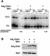
Fig. 5. Chd1 association with TEF2 is partially dependent upon Rtf1. (A) ChIP analysis of HA3-Chd1 on the TEF2 gene in strains lacking (Δ) or containing (+) a functional RTF1 gene. Strains GHY773, KY620 and FY118 were grown in YPD media and analyzed by ChIP using anti-HA1 antibody and the TEF2 internal primer pair 5 (see Figure 4A). FY118 contains an untagged CHD1 gene (–). Sample preparation, PCR analysis and quantitation were performed as described in Figure 3. (B) Immunoblot analysis of HA3-Chd1 levels in yeast extracts (100 µg) prepared from the same strains used in (A). The filter in the upper panel was stripped and probed with anti-Sse1 antibody to serve as a loading control.

Similar articles

Cited by

References

    1. Alén C., Kent,N.A., Jones,H.S., O’Sullivan,J., Aranda,A. and Proudfoot,N.J. (2002) A role for chromatin remodeling in transcriptional termination by RNA polymerase II. Mol. Cell, 10, 1441–1452. - PubMed
    1. Andrulis E.D., Guzman,E., Doring,P., Werner,J. and Lis,J.T. (2000) High-resolution localization of Drosophila Spt5 and Spt6 at heat shock genes in vivo: roles in promoter proximal pausing and transcription elongation. Genes Dev., 14, 2635–2649. - PMC - PubMed
    1. Bertram M.J. and Pereira-Smith,O.M. (2001) Conservation of the MORF4 related gene family: identification of a new chromo domain subfamily and novel protein motif. Gene, 266, 111–121. - PubMed
    1. Bouazoune K., Mitterweger,A., Langst,G., Imhof,A., Akhtar,A., Becker,P.B. and Brehm,A. (2002) The dMi-2 chromodomains are DNA binding modules important for ATP-dependent nucleosome mobilization. EMBO J., 21, 2430–2440. - PMC - PubMed
    1. Bourgeois C.F., Kim,Y.K., Churcher,M.J., West,M.J. and Karn,J. (2002) Spt5 cooperates with human immunodeficiency virus type 1 Tat by preventing premature RNA release at terminator sequences. Mol. Cell. Biol., 22, 1079–1093. - PMC - PubMed

Publication types

MeSH terms