. 2003 Apr 15;22(8):1878-88.
doi: 10.1093/emboj/cdg187.

Modification of Sm small nuclear RNAs occurs in the nucleoplasmic Cajal body following import from the cytoplasm

Affiliations

Modification of Sm small nuclear RNAs occurs in the nucleoplasmic Cajal body following import from the cytoplasm

Beáta E Jády et al. EMBO J. .

Abstract

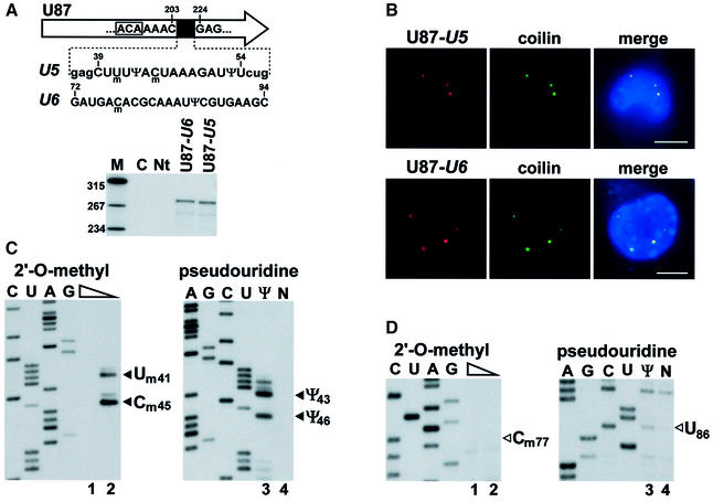
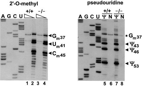
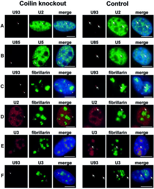
Biogenesis of functional spliceosomal small nuclear RNAs (snRNAs) includes the post-transcriptional covalent modification of numerous internal nucleotides. We have recently demonstrated that synthesis of 2'-O-methylated nucleotides and pseudouridines in the RNA polymerase II-synthesized Sm snRNAs is directed by sequence-specific guide RNAs. Here, we provide evidence supporting the notion that modification of Sm snRNAs occurs in nucleoplasmic Cajal bodies (CBs), where modification guide RNAs accumulate. We show that short fragments of Sm snRNAs are correctly and efficiently modified when targeted to CBs, but not when these same fragments are targeted to the nucleolus. We also demonstrate that internal modification of the U2 snRNA occurs exclusively after nuclear import of the newly assembled Sm snRNP from the cytoplasm. Finally, we show that p80 coilin, the CB marker protein, is not required for snRNA modification. In coilin knockout cells, Sm snRNAs and their modification guide RNAs colocalize in residual CBs, which do not stockpile fibrillarin and fail to recruit the U3 small nucleolar RNA.


Figures

None
Fig. 1. Modification of Sm mutant pre-U2 snRNA. (A) Human pre-U2 snRNA with an altered Sm binding site accumulates in the cytoplasm. Simian COS-7 cells were transfected with the pU2sm expression construct. RNAs isolated from transfected cells (T) or from the nuclear (Nu), nucleoplasmic (Np), nucleolar (No) or cytoplasmic (Cy) fractions of transfected cells were analysed by RNase A/T1 mapping. Protected RNAs corresponding to the 3′ end-extended precursor of U2sm and the endogenous U2 snRNA (U2endo) are indicated. Note that the exposure time of the upper panel was about 50 times longer than that of the lower panel. Lane C, control mapping with Escherichia coli tRNA. Lane M, molecular markers. (B) Primer extension mapping of 2′-O-methylated nucleotides. RNAs isolated from the cytoplasmic (lanes 1 and 2) or nuclear (lanes 3 and 4) fraction of COS-7 cells expressing the pre-U2sm RNA were annealed with terminally labelled oligonucleotides complementary to the mutant (lanes 1 and 2) or wild-type (lanes 3 and 4) U2 RNA and incubated with AMV reverse transcriptase in the presence of 1 mM (lanes 1 and 3) or 0.004 mM (lanes 2 and 4) dNTPs. The extended products were fractionated on a 6% denaturing polyacrylamide gel. Lanes C, U, A and G represent dideoxy sequencing reactions performed on the pU2sm and pU2 plasmids. Stop signals corresponding to 2′-O-methylated nucleotides and expected positions of 2′-O-methylated nucleotides are indicated by closed and open arrowheads, respectively. (C) Mapping of pseudouridine residues in the U2sm RNA. Cytoplasmic (lanes 1 and 2) and nuclear (lanes 3 and 4) RNAs, either treated (Ψ) or non-treated (N) with CMC, were analysed by primer extension. The observed and expected positions of pseudouridines are indicated by closed and open arrowheads, respectively. Asterisks indicate stops generated by 2′-O-methylated nucleotides. For other details, see the legend to (B).
None
Fig. 2. Mapping of 2′-O-methylated nucleotides and pseudouridines in human pre-U2 snRNA. A terminally labelled oligonucleotide (O16) was annealed to HeLa cytoplasmic RNA or in vitro transcribed pre-U2 snRNA. RNA samples used for pseudouridylation mapping had been treated with CMC and alkali buffer (lanes 1 and 5) or only with alkali buffer (lanes 2 and 6). Primer extension was performed with AMV reverse transcriptase in the presence of 1 mM (lanes 1, 2, 3, 5, 6 and 7) or 0.004 mM (lanes 4 and 8) dNTPs. Lanes A, G, C and U, dideoxy sequencing reactions. The expected positions of 2′-O-methylated nucleotides and pseudouridines are indicated by open arrows and arrowheads, respectively. For other details, see the legend to Figure 1.
None
Fig. 3. Nucleolar expression and modification of mouse ribosomal minigene transcripts tagged with human U5, U6 and 5.8S-specific sequences. (A) Schematic structure of the pW-U5, pW-U6 and pW-5.8S expression constructs and in situ localization of the expressed W-U5 RNA in mouse L929 cells. The mouse pol I promoter and terminator (term), fragments derived from the 5′ (hatched boxes) and 3′ (open box) external transcribed spacers (ETS) of the mouse rRNA gene, and a fragment of the chloramphenicol acetyltransferase (CAT) gene are shown. Insertion of synthetic DNAs into the XbaI (Xb) and XhoI (Xh) sites of pW resulted in pW-U5, pW-U6 and pW-5.8S. Nucleotides 2′-O-methylated (m) and pseudouridylated (Ψ) in the human U5, U6 and 5.8S RNAs are indicated. Mouse cells transfected with the pW-U5 expression construct were hybridized with a fluorescent oligonucleotide probe specific for the W-U5 transcript. The nucleolus was visualized by expression of GFP-tagged fibrillarin. Nuclear DNA was stained with DAPI (blue). Bar, 10 µm. (B) Mapping of 2′-O-methylated nucleotides and pseudouridines. Mouse L929 cells were transfected with the pW-U5, pW-U6 or pW-5.8S expression construct as indicated. RNAs isolated from transfected (Tr) or non-transfected (Nt) cells were analysed by primer extension in the presence of 1 mM (lanes 1, 3, 5 and 6) or 0.004 mM (lanes 2 and 4) dNTPs. RNA analysed on lane 5 was treated with CMC. Lanes C, U, G and A represent dideoxy sequencing reactions performed on the pW-U5, pW-U6 or pW-5.8S plasmid. Stops representing modified nucleotides are indicated by closed arrowheads. The expected positions of 2′-O-methylated nucleotides and pseudouridines in the W-U5 transcript are indicated by open arrowheads.
None
Fig. 4. Restoration of 2′-O-methylation of the W-U5 RNA. (A) Schematic structure of the pCMV-globin-yU24m expression construct and expression of the yU24m snoRNA. The promoters of the cytomegalovirus (CMV) and the SP6 RNA polymerase, the exons of the human β-globin gene (E1–E3), the polyadenylation region of the bovine growth hormone gene (PA) are shown. The coding region of a mutant version of the yeast U24 snoRNA (yU24m) is represented by an open arrow. The sequence of the D box and the altered target recognition region of the yU24m snoRNA (in italics) positioning the C45 residue (circled) in the U5 snRNA for 2′-O-methylation are shown. Relevant restriction sites are indicated (H, HindIII; C, ClaI; X, XhoI). RNase A/T1 mapping of RNAs obtained from mouse L929 cells either transfected (T) or non-transfected (N) with the pCMV-globin-yU24m construct. The protected RNA triplet likely reflects a heterogeneity of the 3′ terminus of the expressed yU24m snoRNA (Ganot et al., 1997b). Lane C, control mapping with E.coli tRNA. Lane M, size markers. (BIn situ localization of yU24m RNA. Mouse L929 cells co-transfected with pW-U5, pCMV-globin-yU24m and pfibrillarin-GFP were probed with a fluorescent oligonucleotide complementary to the yU24m RNA. Bar, 10 µm. (C) Primer extension mapping of 2′-O-methylated nucleotides. RNA isolated from mouse cells co-expressing the yU24m and W-U5 RNAs was mapped with a primer specific for the W-U5 RNA. For other details, see the legend to Figure 1.
None
Fig. 5. Modification of a U5-specific sequence in the CB. (A) Expression of U87 scaRNA tagged with U5- or U6-specific sequences. A fragment of the human U87 scaRNA gene between C203 and G224 was replaced for short regions of the U5 or U6 snRNAs. The ACA motif of U87 is boxed. For a detailed structure of the U87 RNA, see figure 1 in Darzacq et al. (2002). Nucleotides facilitating cloning are in lower case letters. Pseudouridines (Ψ) and 2′-O-methylated nucleotides (m) are shown. The U87-U5 and U87-U6 genes were inserted into the pCMV-globin expression vector and transfected into mouse L929 cells. RNAs from transfected (U87-U5 and U87-U6) or non-transfected (Nt) cells were analysed by RNase A/T1 mapping. For other details, see the legend to Figure 1A. (BIn situ localization of U87-U5 and U87-U6 RNAs. Mouse cells expressing the U87-U5 or U87-U6 RNAs were hybridized with specific fluorescent oligonucleotides. CBs were visualized by co-expression of RFP–coilin. Bar, 10 µm. (C) Mapping of 2′-O-methylated nucleotides and pseudouridines in the U87-U5 RNA. RNA isolated from mouse cells expressing the U87-U5 RNA was analysed by primer extension. (D) Primer extension mapping of 2′-O-methylated nucleotides and pseudouridines in the U87-U6 RNA. For other details, see the legend to Figure 3B.
None
Fig. 6. The U5 snRNA is correctly modified in coilin knockout mouse embryonic fibroblast cells. RNAs from coilin knockout (–/–) or control (+/+) cells were subjected to primer extension analysis with a U5-specific primer. For other details, see the legend to Figure 1B and C.
None
Fig. 7. Identification of a novel CB-like structure accumulating scaRNAs and spliceosomal snRNAs. MEF cells derived from coilin knockout or control mice were transfected with the pCMV-globin-U93 (A, C and F), pCMV-globin-U85 (B) and/or pfibrillarin-GFP (C, D and E) expression constructs. Distribution of the U93, U85, U2 and U5 RNAs was detected by in situ hybridization with sequence-specific fluorescent oligonucleotide probes. Extranucleolar foci corresponding to canonical CBs (arrows) and ‘residual CBs’ containing fibrillarin and U3 snoRNA (open arrowheads) or scaRNAs and snRNAs (closed arrowheads) are indicated. DAPI staining of nuclear DNA was omitted in (D). Bar, 10 µm.

Similar articles

Cited by

References

    1. Almeida F., Saffrich,R., Ansorge,W. and Carmo-Fonseca,M. (1998) Microinjection of anti-coilin antibodies affects the structure of coiled bodies. J. Cell Biol., 142, 899–912. - PMC - PubMed
    1. Andrade L.E., Chan,E.K., Raska,I., Peebles,C.L., Roos,G. and Tan,E.M. (1991) Human autoantibody to a novel protein of the nuclear coiled body: immunological characterization and cDNA cloning of p80-coilin. J. Exp. Med., 173, 1407–1419. - PMC - PubMed
    1. Ares M. Jr, Mangin,M. and Weiner,A.M. (1985) Orientation-dependent transcriptional activator upstream of a human U2 snRNA gene. Mol. Cell. Biol., 5, 1560–1570. - PMC - PubMed
    1. Bakin A. and Ofengand,J. (1993) Four newly located pseudouridylate residues in Escherichia coli 23S ribosomal RNA are all at the peptidyltransferase center: analysis by the application of a new sequencing technique. Biochemistry, 32, 9754–9762. - PubMed
    1. Bauer D.W. and Gall,J.G. (1997) Coiled bodies without coilin. Mol. Biol. Cell, 8, 73–82. - PMC - PubMed

Publication types