. 2003 Apr 15;22(8):1909-21.
doi: 10.1093/emboj/cdg175.

Characterization of a novel RNA-binding region of eIF4GI critical for ribosomal scanning

Affiliations

Characterization of a novel RNA-binding region of eIF4GI critical for ribosomal scanning

Déborah Prévôt et al. EMBO J. .

Abstract

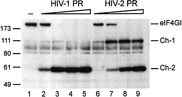
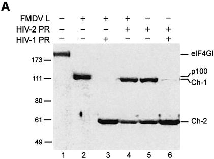
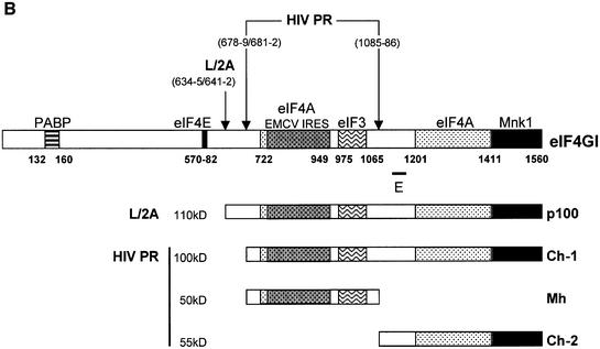
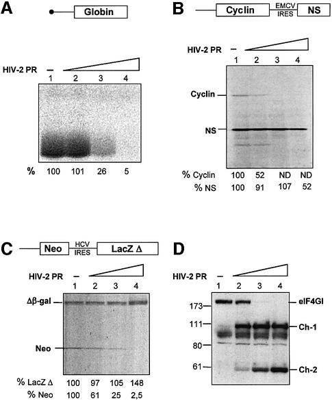
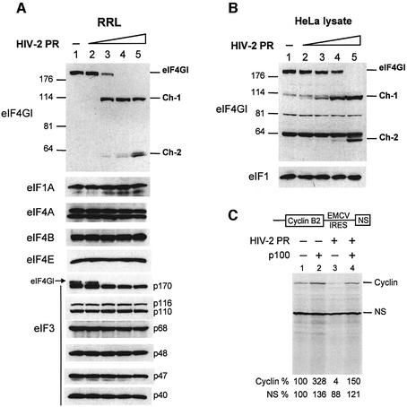
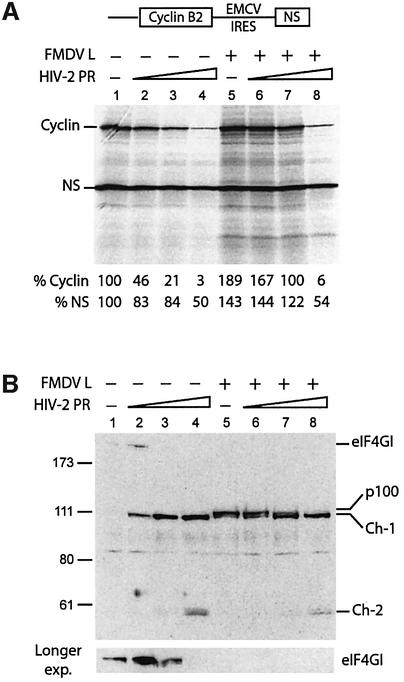
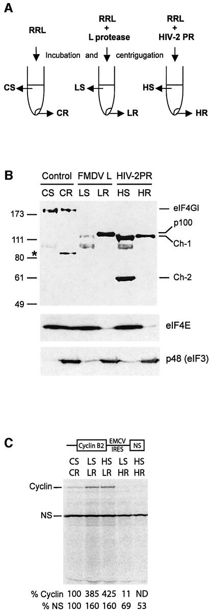
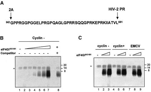
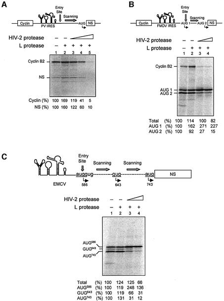
The eukaryotic translation initiation factor eIF4GI binds several proteins and acts as a scaffold to promote preinitiation complex formation on the mRNA molecule (48S). Following mRNA attachment this complex scans along the messenger in a 5' to 3' direction until it locates and recognizes the initiation start codon. By using a combination of retroviral and picornaviral proteases (HIV-2 and L respectively) in the reticulocyte lysate system, we have characterized a 40 amino acid (aa) region of eIF4GI (aa 642-681) that exhibits general RNA-binding properties. Removal of this domain by proteolytic processing followed by translational assays showed virtually no inhibition of internal ribosome entry on the encephalomyocarditis virus, but resulted in drastic impairment of ribosome scanning as demonstrated by studying poliovirus and foot-and-mouth disease virus translation. Based on these findings, we propose that this 40 aa motif of eIF4GI is critical for ribosome scanning.


Figures

None
Fig. 1. eIF4GI is cleaved by recombinant HIV-1 and HIV-2 proteases. RRL (10 µl) was incubated with buffer (lane 1) or increasing amounts of recombinant HIV-1 PR (lanes 2–5: 0.25, 1.25, 5 and 12.5 ng/µl) or HIV-2 PR (lanes 6–9: 0.5, 2.5, 10 and 25 ng/µl) for 1 h at 30°C in a final volume of 20 µl. A 1 µl aliquot was resolved on 10% SDS–PAGE, proteins transferred to PVDF and the membrane was incubated with antibodies specific to the C-terminal part of eIF4GI (serum E, see Figure 2B). The resulting fragments and molecular weight markers (in kDa) are indicated on the figure.
None
Fig. 2. Characterization of the C-terminal cleavage sites of eIF4GI. (A) RRL (10 µl) was incubated without (lane 1) or with L protease (1 µl, lanes 2–4) or HIV-2 PR (10 ng/µl, lanes 5 and 6), for 1 h at 30°C in a final volume of 20 µl. HIV-2 PR (20 ng/µl; lane 4) or 10 ng/µl HIV-1 PR (lanes 3 and 6) were then added and the mixture further incubated for 1 h. The samples were analysed by SDS–PAGE and western blotting as described in Figure 1. (B) The eIF4GI molecule is schematically represented with its different interaction domains for PABP and eIF4E (Gingras et al., 1999b), eIF4A (Lomakin et al., 2000; Morino et al., 2000), eIF3 (Korneeva et al., 2000), Mnk-1 (Morino et al., 2000) and the EMCV IRES (Lomakin et al., 2000). Cleavage sites for L/2A and HIV proteases, the obtained C-terminal fragments and the E epitope used for the western blot analysis are represented.
None
Fig. 3. HIV-2 PR abolishes capped and uncapped mRNAs translation, but moderately inhibits EMCV IRES-driven translation. (A, B and C) A RRL under full translation conditions (see Materials and methods) was pre-incubated without (lanes 1) or with different amounts of HIV-2 PR (lanes 2–4: 3.5, 7 and 14 ng/µl). After 1 h at 30°C, Palinavir (10 µM final concentration) was added to the reactions. Different transcripts (schematically represented on the upper part of each panel) including (A) natural capped globin mRNA (2.5 ng/µl), (B) XL–EMCV mRNA (10 ng/µl) and (C) DC–HCV mRNA (10 ng/µl) were translated under these conditions. Samples were processed on 15% SDS–PAGE, submitted to autoradiography, the relative intensities of the bands were quantified and the results are presented at the bottom of each panel. (D) Aliquots of the samples 1–4 from (A) were resolved on 10% SDS–PAGE, proteins transferred to PVDF and the membranes were incubated with antibodies specific to the C-terminal part of eIF4GI (serum E). The resulting fragments and molecular weight markers (in kDa) are indicated on the figure.
None
Fig. 4. Inhibition of translation observed with HIV-2 PR is due to cleavage of eIF4GI (A and B). (A) RRL (10 µl) or (B) HeLa lysate (127.5 µg) was incubated with buffer (lane 1) or increasing amounts of recombinant HIV-2 PR (lanes 2–5: 2.5, 5, 10 and 25 ng/µl) for 1 h at 30°C in a final volume of 20 µl. Aliquots were resolved on SDS–PAGE, proteins transferred to PVDF and the membranes were incubated with antibodies specific to eIF1, eIF1A, eIF3, eIF4A, eIF4B, eIF4E and the C-terminal part of eIF4GI, as indicated on the left side of each panel. For eIF4GI, resulting fragments and molecular weight markers (in kDa) are indicated on the figure. (C) RRL under full translation conditions was pre-incubated without (lanes 1 and 2) or with (lanes 3 and 4) 7 ng/µl HIV-2 PR. After 30 min at 30°C, Palinavir (10 µM final concentration) was added to the reactions with 1 ng/µl recombinant p100 fragment (lanes 2 and 4) or buffer (lanes 1 and 3). Uncapped XL–EMCV mRNA (10 ng/µl) was then translated under these conditions. Samples were processed on 15% SDS–PAGE, submitted to autoradiography, the relative intensities of the bands were quantified and the results are presented at the bottom of the figure.
None
Fig. 5. ‘Clipping’ of p100 by HIV-2 PR inhibits uncapped translation. (A) RRL under full translation conditions was pre-incubated without (lanes 1–4) or with (lanes 5–8) 0.5 µl of L protease for 20 min at 30°C. Either buffer (lanes 1 and 5) or 1.75 ng/µl (lanes 2 and 6), 3.5 ng/µl (lanes 3 and 7) and 7 ng/µl (lanes 4 and 8) of HIV-2 PR were then added to the reactions. After 1 h at 30°C, Palinavir (10 µM final concentration) was added and translation of XL–EMCV mRNA (10 ng/µl) was performed under these conditions. Samples were analysed as described in Figure 5. (B) Western blot analysis of samples 1–8 of (A) was performed as described in Figure 1. A longer exposition of the upper part of the blot, corresponding to proteins of high molecular weight including intact eIF4GI is presented at the bottom of the panel.
None
Fig. 6. Ch-1 fragment can support IRES-mediated, but not uncapped mRNA translation. (A) Schematic representation of RRL incubation and fractionation. (B) RRL (200 µl) was incubated alone, with 15 µl of in vitro translated L protease or with 1.4 µg of HIV-2 PR. After 1 h at 30°C, L and HIV-2 proteases were inhibited by 0.5 mM E-64 or 10 µM Palinavir, respectively, and lysates were ultracentrifuged [see Materials and methods and (A)]. Aliquots of lysate corresponding to 2 µl of parent RRL were resolved on a 10% SDS–PAGE, and western blot analysis was performed with antibodies specific to the C-terminal part of eIF4GI (upper), eIF4E (middle) and p48 protein of eIF3 (bottom). The resulting proteins or fragments observed, and molecular weight markers (in kDa) are indicated on the figure. The asterisk denotes a non-specific reaction with the eIF4GI antibody. CS, supernatant control; CR, ribosomes control; LS, L-treated supernatant; LR, L-treated ribosomes; HS, HIV-2-treated supernatant; HR, HIV-2-treated ribosomes. (C) Different reconstituted lysates were obtained by combining 1 µl of ribosomes and 5 µl of supernatants described in (A). XL–EMCV mRNA (10 ng/µl) was then translated under these conditions. Samples were analysed as described in Figure 3.
None
Fig. 7. eIF4GI642–681 can be crosslinked to various mRNAs. (A) Sequence of the 40 aa peptide (eIF4GI642–681), the 2A and HIV-2 cleavage sites are indicated on the figure. (B) Increasing amounts of chemically synthesized eIF4GI642–681 (lane 1, 0 µg; lane 2, 0.015 µg; lane 3, 0.030 µg; lane 4, 0.06 µg; lane 5, 0.12 µg; lane 6, 0.24 µg; lanes 7 and 8, 0.48 µg) was used in crosslinking assays with 32P-labelled uncapped cyclin probe (0.2 pmol; 75 000 c.p.m.) in the absence (lanes 1–7) or presence of unlabelled luciferase competitor (lane  8, 7 pmol). Following RNase A treatment, proteins were separated on 20% SDS–PAGE and the dried gel was exposed to autoradiography. The asterisk denotes a non-specific band which probably results from incomplete digestion of the radiolabelled transcript. (C) Crosslinking assays were performed in the absence (lanes 1, 4 and 7) or presence of chemically synthesized eIF4GI642–681 (lanes 2, 5 and 8, 0.25 µg; lanes 3, 6 and 9, 0.75 µg) with various 32P-labelled probes (75 000 c.p.m.): uncapped cyclin (lanes 1–3), capped cyclin (lanes 4–6) and uncapped EMCV (lanes 7–9). Following RNase A treatment, proteins were separated on 20% SDS–PAGE and the dried gel was subjected to autoradiography. Position of molecular weight markers are indicated on the right-hand side of the figure in kDa.
None
Fig. 8. Conversion of p100 into Ch-1 inhibits ribosomal scanning. RRL under full translation conditions was pre-incubated without (lanes 1) or with (lanes 2–5) 0.5 µl of L protease for 20 min at 30°C. Either buffer (lanes 1 and 2) or 3.5 ng/µl (lanes 3), 7 ng/µl (lanes 4) or 14 ng/µl (A, lane 5) of HIV-2 PR was added to the reaction. After 1 h at 30°C, Palinavir (10 µM final) was added and translation was programmed with (A) bicistronic XL–PV mRNA (10 ng/µl), (B) bicistronic XL–FMDV mRNA (10 ng/µl) or (C) monocistronic EMCV–PV (10 ng/µl). Samples were processed on SDS–PAGE, submitted to autoradiography and the relative intensities of the bands were quantified and the results are presented at the bottom of each panel. They are expressed as a percentage of total initiation events (Total), or as the relative translation efficiency for each individual initiation codon as indicated on the figure. A schematic diagram of the constructs used and their initiation sites are represented on top of each panel.

Similar articles

Cited by

References

    1. Asano K., Clayton,J., Shalev,A. and Hinnebusch,A.G. (2000) A multifactor complex of eukaryotic initiation factors, eIF1, eIF2, eIF3, eIF5 and initiator tRNA(Met) is an important translation initiation intermediate in vivo. Genes Dev., 14, 2534–2546. - PMC - PubMed
    1. Belsham G.J. (1992) Dual initiation sites of protein synthesis on foot-and-mouth disease virus RNA are selected following internal entry and scanning of ribosomes in vivo. EMBO J., 11, 1105–1110. - PMC - PubMed
    1. Belsham G.J., McInerney,G.M. and Ross-Smith,N. (2000) Foot-and-mouth disease virus 3C protease induces cleavage of translation initiation factors eIF4A and eIF4G within infected cells. J. Virol., 74, 272–280. - PMC - PubMed
    1. Berlioz C. and Darlix,J.L. (1995) An internal ribosomal entry mechanism promotes translation of murine leukemia virus gag polyprotein precursors. J. Virol., 69, 2214–2222. - PMC - PubMed
    1. Borman A.M., Kirchweger,R., Ziegler,E., Rhoads,R.E., Skern,T. and Kean,K.M. (1997) elF4G and its proteolytic cleavage products: effect on initiation of protein synthesis from capped, uncapped and IRES-containing mRNAs. RNA, 3, 186–196. - PMC - PubMed

Publication types