. 2003 Apr 14;161(1):119-29.
doi: 10.1083/jcb.200212174. Epub 2003 Apr 7.

GDNF promotes tubulogenesis of GFRalpha1-expressing MDCK cells by Src-mediated phosphorylation of Met receptor tyrosine kinase

Affiliations

GDNF promotes tubulogenesis of GFRalpha1-expressing MDCK cells by Src-mediated phosphorylation of Met receptor tyrosine kinase

Anna Popsueva et al. J Cell Biol. .

Abstract

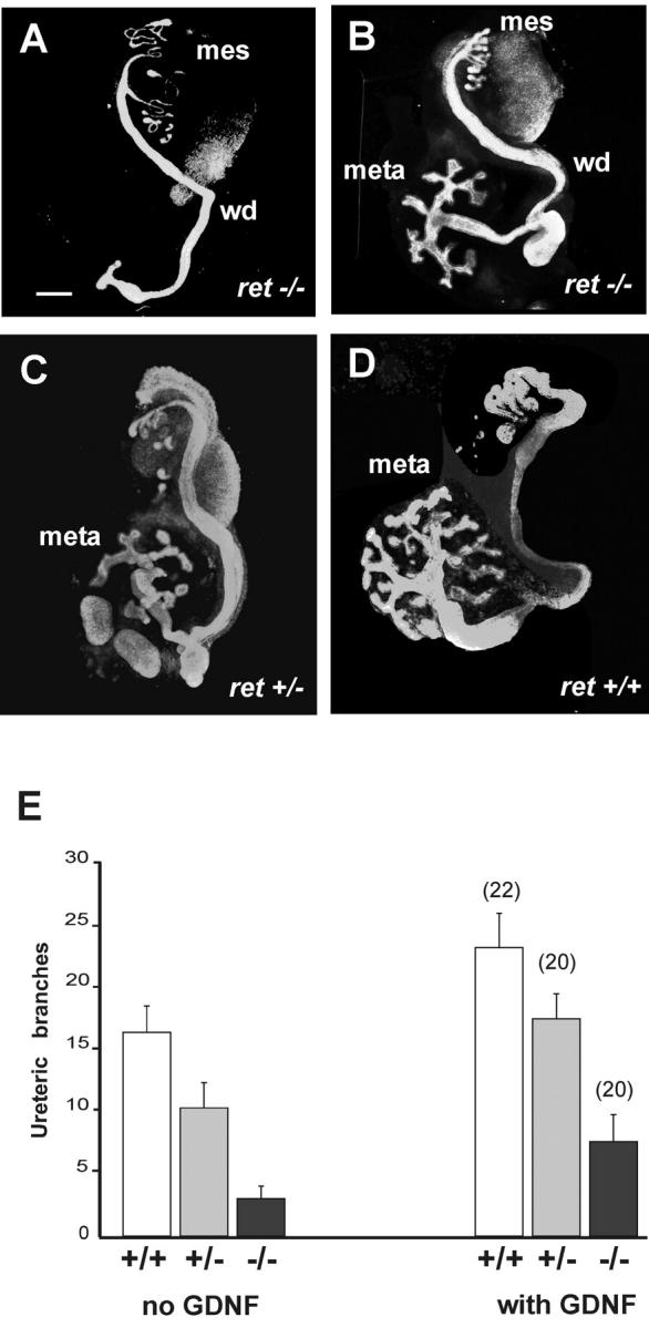
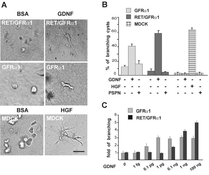
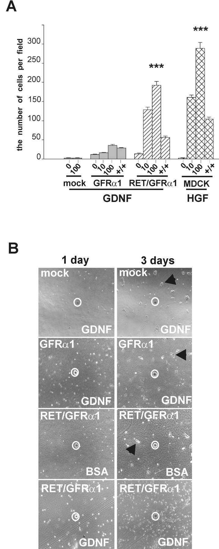
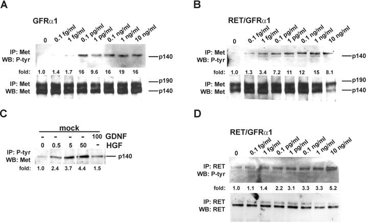
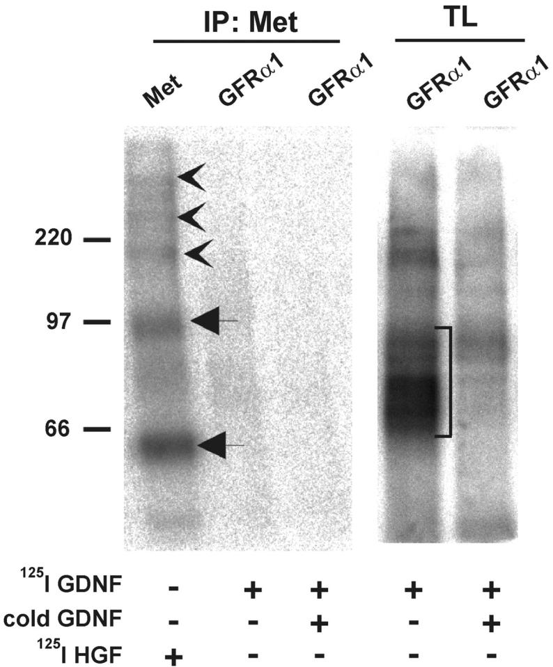
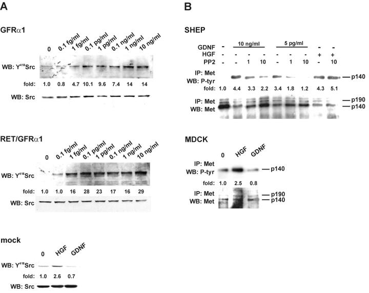
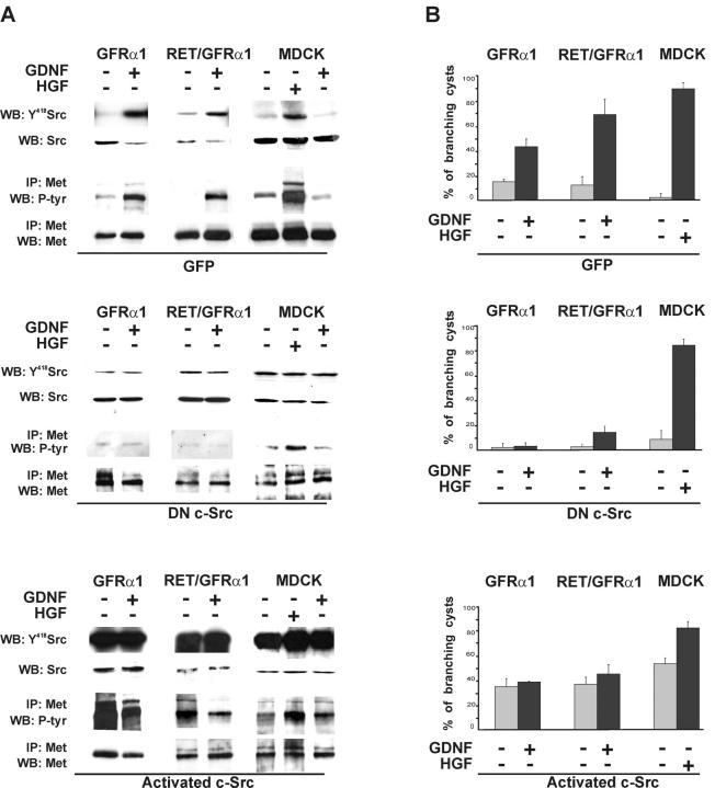
Glial cell line-derived neurotrophic factor (GDNF) and hepatocyte growth factor (HGF) are multifunctional signaling molecules in embryogenesis. HGF binds to and activates Met receptor tyrosine kinase. The signaling receptor complex for GDNF typically includes both GDNF family receptor alpha1 (GFRalpha1) and Ret receptor tyrosine kinase. GDNF can also signal independently of Ret via GFRalpha1, although the mechanism has remained unclear. We now show that GDNF partially restores ureteric branching morphogenesis in ret-deficient mice with severe renal hypodysplasia. The mechanism of Ret-independent effect of GDNF was therefore studied by the MDCK cell model. In MDCK cells expressing GFRalpha1 but no Ret, GDNF stimulates branching but not chemotactic migration, whereas both branching and chemotaxis are promoted by GDNF in the cells coexpressing Ret and GFRalpha1, mimicking HGF/Met responses in wild-type MDCK cells. Indeed, GDNF induces Met phosphorylation in several ret-deficient/GFRalpha1-positive and GFRalpha1/Ret-coexpressing cell lines. However, GDNF does not immunoprecipite Met, making a direct interaction between GDNF and Met highly improbable. Met activation is mediated by Src family kinases. The GDNF-induced branching of MDCK cells requires Src activation, whereas the HGF-induced branching does not. Our data show a mechanism for the GDNF-induced branching morphogenesis in non-Ret signaling.


Figures

Figure 1.
Figure 1.
Exogenous GDNF partially restores ureteric branching of ret-deficient kidneys. (A–D) Urogenital block explants including the Wolffian duct (wd), mesonephros (meso), and metanephros (meta) from E11 ret −/− (A and B), ret +/− (C), and ret +/+ (D) mouse embryos. The urogenital blocks from each embryo were separately cultured for 4 d, one side without GDNF (A, C and D) and the other one with 50 ng/ml of GDNF (B). The explants were fixed and immunolabeled as whole mounts with pan-cytokeratin antibodies. Bar, 200 μm. (E) The number of ureteric branches of ret −/−, ret +/−, and ret +/+ kidneys with or without GDNF supplementation. The results represent the means ± SEM. GDNF significantly increases ureteric branch number in ret −/− explants compared with the control media (P < 0.01).
Figure 2.
Figure 2.
GDNF induces branching of GFRα1-expressing MDCK cells in three-dimensional collagen gel. (A) Ret/GFRα1- and GFRα1-expressing MDCK cells were grown in collagen gel with GDNF (100 ng/ml), and wild-type MDCK cells were grown in collagen gel with HGF (50 ng/ml). BSA (100 ng/ml) was used as a negative control. Bar, 100 μm. (B) GDNF induces branching of GFRα1 and Ret/GFRα1 cells but not wild-type MDCK cells, which only respond to HGF. Persephin (PSPN; 100 ng/ml) does not induce branching of any MDCK cell line tested. From the total number of cysts in the field, the percentage of cysts with long branches was calculated. Only the branches with the length of more than two cyst diameters were counted. (C) Dose dependency of the GDNF-induced branching of GFRα1- and Ret/GFRα1-expressing MDCK cells. GDNF concentrations are marked per ml. Results are reported as fold of branching cysts over the noninduced control. Means ± SEM of five to eight counted fields are shown. The results are representative of five (A and B) and three (C) independent experiments. (B and C) GDNF significantly increases branching in GFRα1- and Ret/GFRα1-expressing MDCK and HGF increases branching of wild-type MDCK (B) compared with the control media (P < 0.001).
Figure 3.
Figure 3.
GFRα1-expressing, ret -deficient MDCK cells do not show a chemotactic response to GDNF. (A) In the Boyden chamber chemotaxis assay, the mock-transfected, GFRα1, and Ret/GFRα1 cells were exposed to GDNF (10 and 100 ng/ml), and wild-type MDCK were exposed to HGF (10 and 100 ng/ml). The number of cells was counted as described in Materials and methods. +/+, 100 ng/ml of GDNF or 50 ng/ml of HGF were added to both chambers to assay chemokinesis. The results represent the means ± SEM (n = 3). ***P < 0.001. (B) Chemoattraction assay on collagen matrix. Only Ret/GFRα1-expressing cells migrate toward GDNF-soaked beads. BSA-soaked agarose beads were used as negative control. Beads are marked by a white circle. Note that mock, GFRα1-expressing cells with GDNF-soaked beads and Ret/GFRα1-expressing cells with BSA-soaked bead form clusters of adherent cells (marked with arrowhead) after 3 d, whereas the Ret/GFRα1-expressing cells migrating toward the GDNF bead are scattered.
Figure 4.
Figure 4.
GDNF induces phosphorylation of Met. (A and B) Dose-dependent phosphorylation of Met by GDNF in GFRα1- and Ret/GFRα1-expressing MDCK cells. Met was activated in 15 min after GDNF application. The bottom panels show the reprobing of the same filter with anti-Met antibodies. The numbers below the lanes indicate the fold of induction of Met tyrosine kinase. (C) Phosphorylation of Met in mock-transfected MDCK cells. Concentrations of GDNF and HGF are given in ng/ml. 30 μg of total proteins were incubated with 10 μl of immobilized phosphotyrosine mAbs, and immunocomplexes were washed and analyzed as described in Materials and methods. (D) Dose-dependent activation of Ret by GDNF in Ret/GFRα1-expressing MDCK cells. The bottom panel shows the reprobing of the same filter with anti-Ret antibodies. The numbers below the lanes indicate the fold of induction of Ret tyrosine kinase. IP, immunoprecipitation; WB, Western blotting; P-tyr, phosphotyrosine. The results are representative of three independent experiments.
Figure 5.
Figure 5.
GFRα1 does not complex with Met. Binding of 125I-GDNF to COS7 cells transfected with gfrα1 and 125I-HGF to wild-type COS7 followed by cross-linking with EDC together with sulfo-NHS. Immunoprecipitates with anti-Met antibodies (IP:Met) were analyzed by SDS-PAGE under reducing conditions. In total lysates (TL), different complexes of 125I-GDNF (monomers or dimers) and the dimers of GFRα1 are marked with a square bracket. 125I-HGF α subunit and proHGF are marked by arrows. 125I-HGF–Met complexes are indicated by arrowheads. The results are representative of five independent experiments.
Figure 6.
Figure 6.
GDNF-induced activation of Met requires Src kinase. (A) Dose-dependent Src kinase activation by GDNF in GFRα1- and Ret/GFRα1-expressing MDCK cells. The activation of Src-type kinases was observed after 15 min. The concentrations of GDNF are marked. The bottom panel shows mock-transfected MDCK cells induced with 50 ng/ml of HGF and 100 ng/ml of GDNF. The numbers below the lane indicate the fold of increase in phosphorylation of Tyr418 of Src. The bottom panels show the reprobing of the same filter with anti-Src antibodies. The results are representative of three independent experiments. (B) SHEP cells were grown with GDNF (10 ng/ml and 5 pg/ml) or HGF (10 ng/ml) in the presence of PP2 (1 and 10 μM). To exclude a possible cytotoxic effect of the solvent, DMSO was added to the controls. The bottom panel shows the reprobing of the same filter with anti-Met antibodies. Bottom picture demonstrates wild-type MDCK cells induced with 50 ng/ml of HGF and 100 ng/ml of GDNF. Numbers below the lane indicate the fold of induction of Met tyrosine kinase. The results are representative of three independent experiments.
Figure 7.
Figure 7.
GDNF-induced branching tubulogenesis of GFRα1- and Ret/GFRα1-expressing cells require c-Src kinase. GFRα1- and Ret/GFRα1-expressing and wild-type MDCK cells were infected with adenovirus constructs containing DN c-Src, activated c-Src, or adeno-GFP. (A) GDNF-induced Met activation depends on c-Src kinase. 1 d after the adenovirus infection, GFRα1- and Ret/GFRα1-expressing MDCK cells were induced with GDNF (50 ng/ml) and wild-type MDCK also with HGF (50 ng/ml). Aliquots of total cell lysates were immunoblotted with anti-Y418 Src, and the rest of lysates were immunoprecipitated with anti-Met antibodies and immunoblotted using antiphosphotyrosine antibodies. The results are representative of two independent experiments. (B) After infection cells were put in collagen gel culture, GFRα1- and Ret/GFRα1-expressing cells were grown with or without GDNF (50 ng/ml), wild-type MDCK with or without HGF (50 ng/ml). After 3 d, the cells were fixed and counted as described in Materials and methods. The results are representative of three independent experiments.

Similar articles

Cited by

References

    1. Abram, C.L., and S.A. Courtneidge. 2000. Src family tyrosine kinases and growth factor signaling. Exp. Cell Res. 254:1–13. - PubMed
    1. Airaksinen, M.S., A. Titievsky, and M. Saarma. 1999. GDNF family neurotrophic factor signaling: four masters, one servant? Mol. Cell. Neurosci. 13:313–325. - PubMed
    1. Baloh, R.H., H. Enomoto, E.M. Johnson, Jr., and J. Milbrandt. 2000. The GDNF family ligands and receptors—implications for neural development. Curr. Opin. Neurobiol. 10:103–110. - PubMed
    1. Behar, T.N., Y.X. Li, H.T. Tran, W. Ma, V. Dunlap, C. Scott, and J.L. Barker. 1996. GABA stimulates chemotaxis and chemokinesis of embryonic cortical neurons via calcium-dependent mechanisms. J. Neurosci. 16:1808–1818. - PMC - PubMed
    1. Behar, T.N., A.E. Schaffner, C.A. Scott, C. O'Connell, and J.L. Barker. 1998. Differential response of cortical plate and ventricular zone cells to GABA as a migration stimulus. J. Neurosci. 18:6378–6387. - PMC - PubMed

Publication types

MeSH terms

Substances