. 2003 Apr 7;197(7):823-9.
doi: 10.1084/jem.20021840.

DC-SIGN (CD209) mediates dengue virus infection of human dendritic cells

Affiliations

DC-SIGN (CD209) mediates dengue virus infection of human dendritic cells

Boonrat Tassaneetrithep et al. J Exp Med. .

Abstract

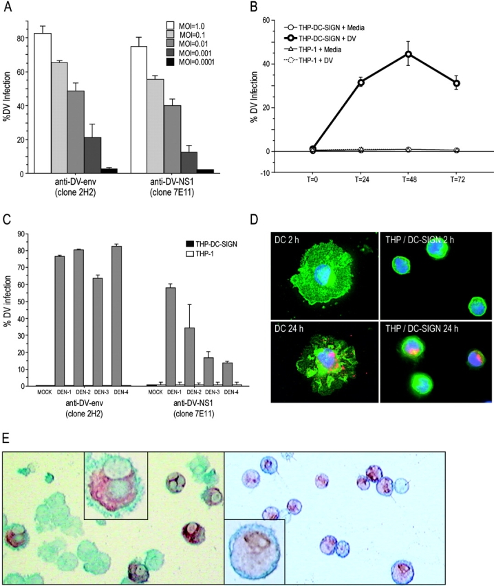
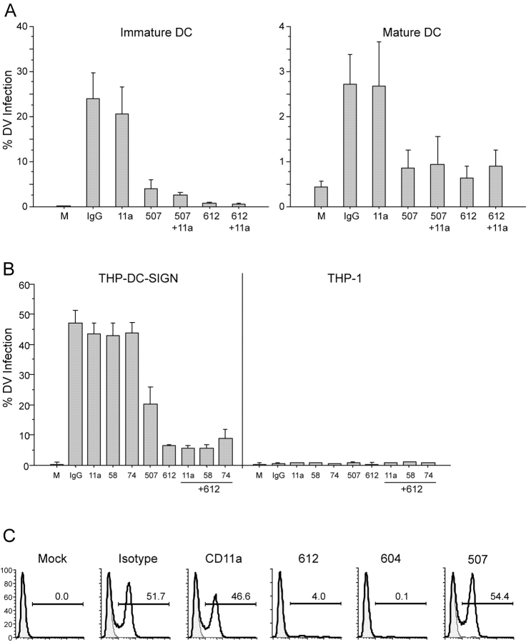
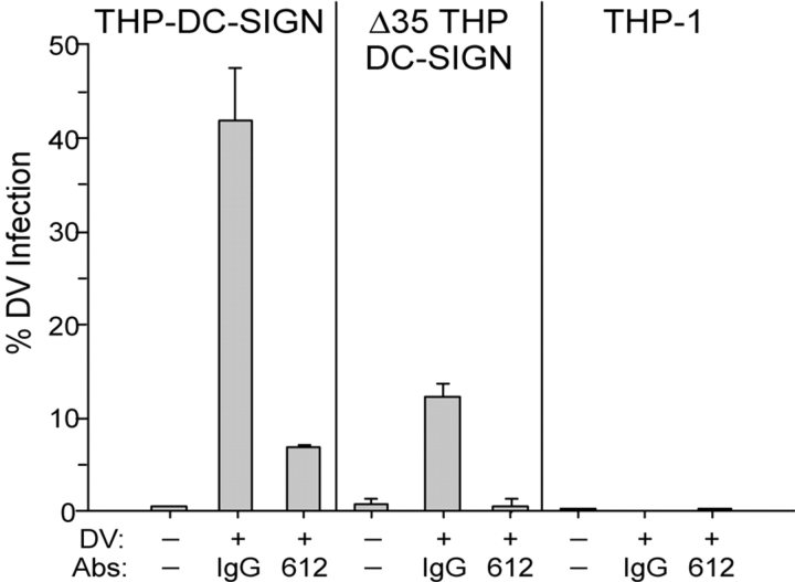
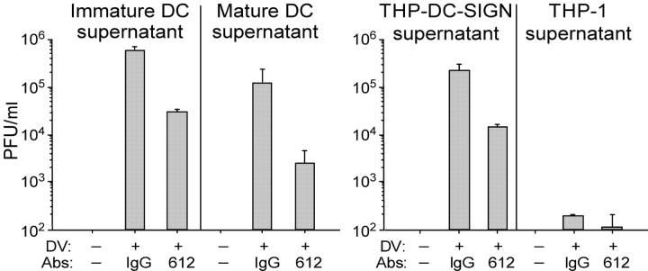
Dengue virus is a single-stranded, enveloped RNA virus that productively infects human dendritic cells (DCs) primarily at the immature stage of their differentiation. We now find that all four serotypes of dengue use DC-SIGN (CD209), a C-type lectin, to infect dendritic cells. THP-1 cells become susceptible to dengue infection after transfection of DC-specific ICAM-3 grabbing nonintegrin (DC-SIGN), or its homologue L-SIGN, whereas the infection of dendritic cells is blocked by anti-DC-SIGN antibodies and not by antibodies to other molecules on these cells. Viruses produced by dendritic cells are infectious for DC-SIGN- and L-SIGN-bearing THP-1 cells and other permissive cell lines. Therefore, DC-SIGN may be considered as a new target for designing therapies that block dengue infection.


Figures

Figure 1.
Figure 1.
Cytofluorometry of DV infection of DC-SIGN–expressing cells. (A) The MFI of DC-SIGN surface expression on DC and THP-1 cells is shown (left). The bars represent means (± SEM) of at least three independent experiments. The top right histogram shows the relative DC-SIGN expression (MFI) on a representative donor's immature (shaded) and MCM-matured DCs (heavy line). The bottom right histogram shows relative DC-SIGN on the surface of THP DC-SIGN (shaded) and THP-1 cells (heavy line). Matched isotype controls are represented by the light dashed line. (B) The mean (± SEM) percentage of DV infection of immature and mature DC (left) and THP DC-SIGN and THP-1 (right) after intracellular staining for dengue envelope antigen (2H2) in at least three independent experiments. (C) Positive correlation between DC-SIGN expression (MFI) on the x axis and percent DV infection as determined by 2H2 binding on the y axis on both DC and THP cells (n = 16).
Figure 2.
Figure 2.
Dose and time dependence of DV infection in THP DC-SIGN cells. (A) Titration of DV2 infection in THP DC-SIGN and THP-1 2 d after infection. The infected cells were stained for DV envelope antigen (clone 2H2) and the NS1 (clone 7E11) and infection was determined by calculating the percentage of fluorescence-positive cells. (B) Kinetics of DV2 infection in THP DC-SIGN and THP-1. The infected cells were harvested at t = 0, 24, 48, and 72 h and stained with 2H2 and 7E11. (C) DV infection rates of all four DV serotypes in THP DC-SIGN and THP-1, using DV 1, 2, 3, and 4. Data are averages of two independent experiments. (D) Representative immunofluorescence experiment at two time points (2 h, top, and 24 h, bottom) showing bound DV envelope complex antigens (red) in DC-SIGN (green)–bearing DC (left) and THP-DC-SIGN (right) cells. Nuclei were labeled with DAPI stain. (E) Representative immunohistochemistry experiment showing bound intracellular DV envelope complex antigens (dark red) in DC-SIGN–bearing cells (blue). The left (immature DC) and right panels (THP DC-SIGN) show cells infected with DV. Original magnifications at 200 (insets, 600).
Figure 3.
Figure 3.
Blocking studies of DV infection in DCs and THP-1. (A) Comparison of percent DV2 infection rate of immature (left) and mature DCs (right) in the presence and absence of anti–DC-SIGN mAbs (clones 120507-specific or 120612–cross-reactive), anti-CD11a, or an irrelevant matched isotype control. (B) Comparison of percent DV2 infection rate of THP DC-SIGN and THP-1 in the presence and absence of specific anti–DC-SIGN mAbs, anti-CD11a, anti-CD58, anti-CD74, or an irrelevant matched isotype control. Data are means (± SEM) of four independent experiments. (C) A representative blocking experiment (one of two) in the THP L-SIGN cells in the presence and absence of specific anti–DC-SIGN or L-SIGN mAbs (120604), anti-CD11a, or an irrelevant matched isotype control.
Figure 4.
Figure 4.
Internalization of DV. Comparison of DV infection of THP DC-SIGN, THP DC-SIGN Δ35 (fully truncated cytoplasmic domain DC-SIGN), and THP-1 in the presence of specific anti–DC-SIGN antibody (clone 120612) or a matched isotype control. Data are the mean (± SEM) of three independent experiments.
Figure 5.
Figure 5.
Plaque assays of cell-free culture supernatants show infectivity. Supernatants transmit DV infection from DV-infected DCs (left) and THP DC-SIGN cells (right) to Vero cells. DCs or THPs were pretreated with an anti–DC-SIGN mAb (clone 120612) or a matched isotype control and exposed to DV for 2 h. Virus and mAb were washed away, and the supernatants were collected 48 h later. Plaque assays were used to determine PFUs in the collected supernatants. Data are the averages of two independent experiments.

Similar articles

Cited by

References

    1. Monath, T.P., and F.X. Heinz. 1996. Flaviviruses. In Fields Virology. B.N. Fields, D.M. Knipe, and P.M. Howley, editors. Lippincott-Raven, Philadelphia. 961–1034.
    1. Gubler, D.J., S. Nalim, R. Tan, H. Saipan, and J. Sulianti Saroso. 1979. Variation in susceptibility to oral infection with dengue viruses among geographic strains of Aedes aegypti. Am. J. Trop. Med. Hyg. 28:1045–1052. - PubMed
    1. Halstead, S.B. 1997. Epidemiology of dengue and dengue hemorrhagic fever. In Dengue and Dengue Hemorrhagic Fever. D.J. Gubler and G. Kuno, editors. Cab International, London. 23–44.
    1. Burke, D.S., A. Nisalak, D.E. Johnson, and R.M. Scott. 1988. A prospective study of dengue infections in Bangkok. Am. J. Trop. Med. Hyg. 38:172–180. - PubMed
    1. Kliks, S.C., A. Nisalak, W.E. Brandt, L. Wahl, and D.S. Burke. 1989. Antibody-dependent enhancement of dengue virus growth in human monocytes as a risk factor for dengue hemorrhagic fever. Am. J. Trop. Med. Hyg. 40:444–451. - PubMed

Publication types

MeSH terms