Comparative Study
. 2003 Apr 15;100(8):4807-12.
doi: 10.1073/pnas.0831079100. Epub 2003 Apr 7.

Plasma membrane localization and function of the estrogen receptor alpha variant (ER46) in human endothelial cells

Affiliations
Comparative Study

Plasma membrane localization and function of the estrogen receptor alpha variant (ER46) in human endothelial cells

Lei Li et al. Proc Natl Acad Sci U S A. .

Abstract

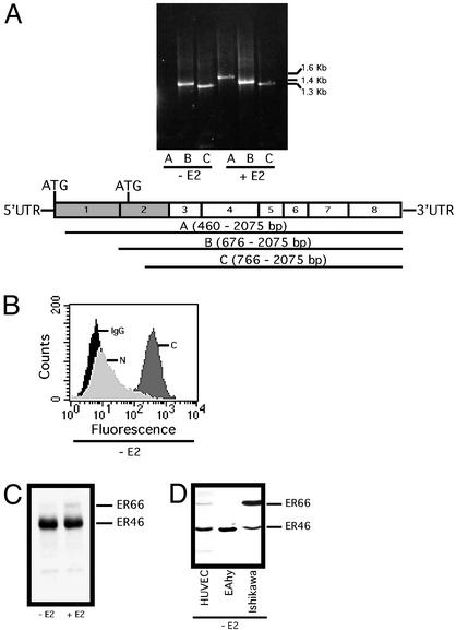
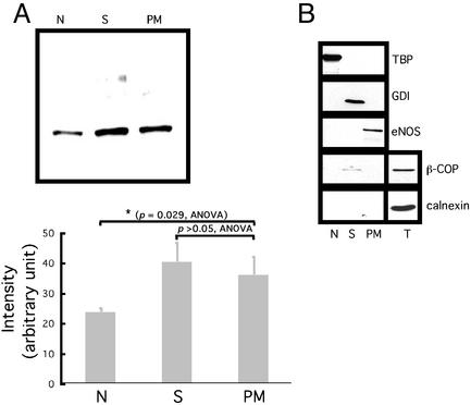
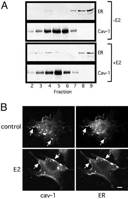
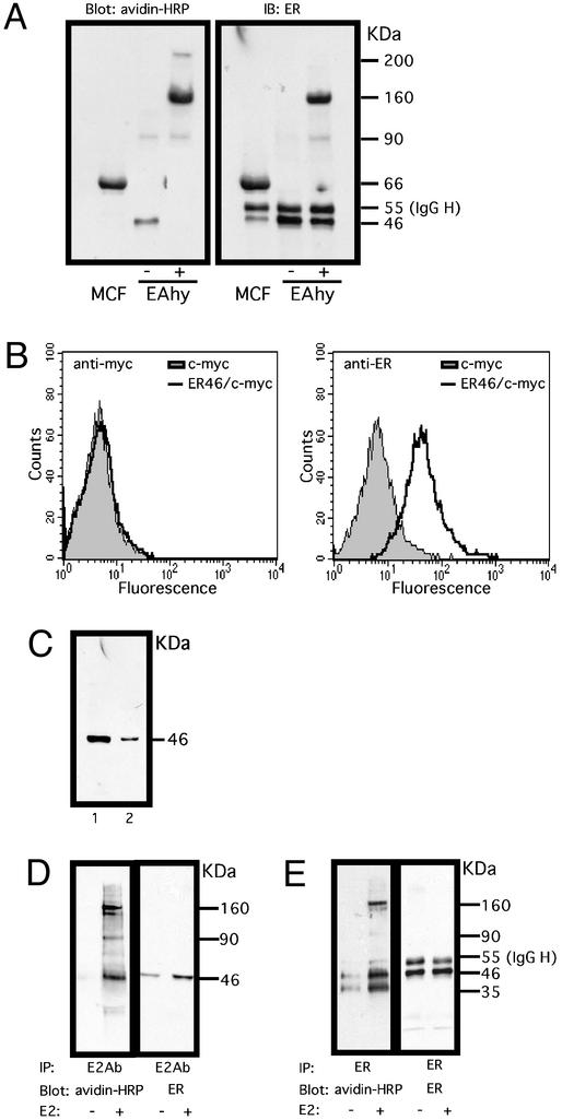
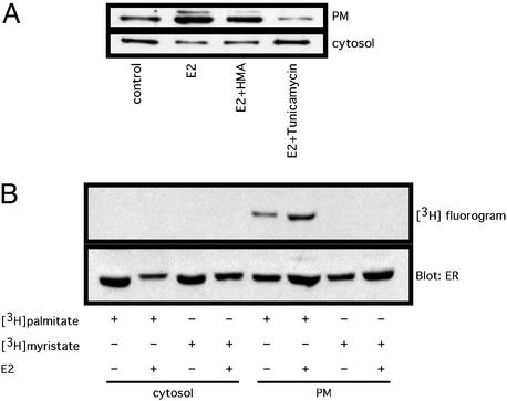
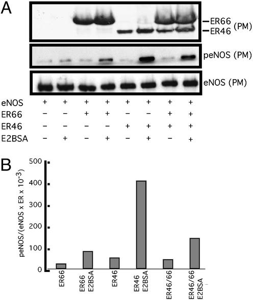
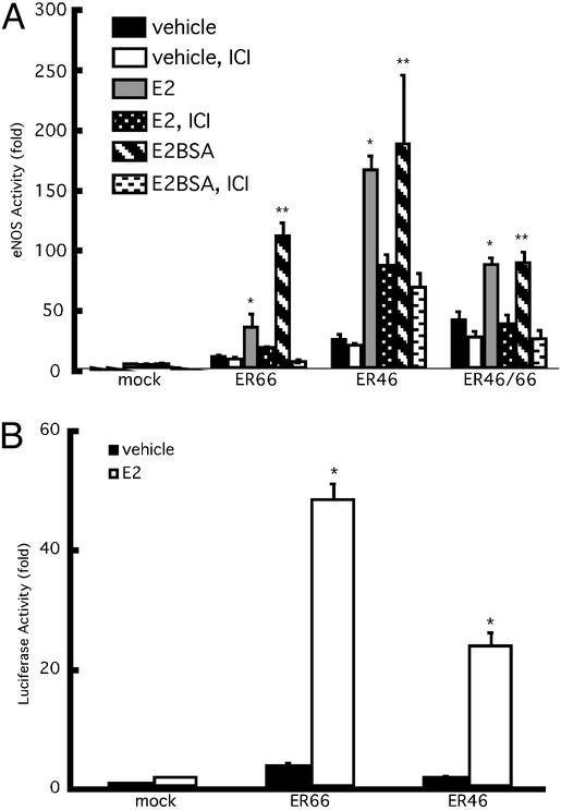
Estrogen receptor (ER) alpha variants have been identified in an array of nonendothelial cells. We previously demonstrated that estrogen rapidly induces nitric oxide release via a phosphatidylinositol 3-kinase/Akt/endothelial nitric-oxide synthase (eNOS) pathway in EA.hy926 cells (immortalized human endothelial cells), which express a 46-kDa ER. We now confirm that, due to alternative splicing, the 46-kDa endothelial cell protein (ER46) is an amino-terminal truncated product of full-length ER alpha (ER66). ER46 is expressed in the plasma membrane, cytosol, and nucleus of resting, estrogen-deprived cells. Flow cytometric and immunofluorescence microscopic analyses demonstrated that the ER46 C but not N terminus is Ab-accessible in the plasma membrane. Inhibition of palmitoylation with tunicamycin and [(3)H]palmitic acid labeling demonstrated an estrogen-induced, palmitoylation-dependent plasma membrane ER46 recruitment, with reorganization into caveolae. In reconstituted, estrogen-stimulated COS-7 (ER-null) cells, membrane ER46 more efficiently triggered membrane eNOS phosphorylation than ER66. Conversely, ER66 more efficiently mediated estrogen response element reporter-gene transactivation than ER46. These results demonstrate that ER46 is localized and further dynamically targeted to the plasma membrane in a palmitoylation-dependent manner. ER46 more efficiently modulates membrane-initiated estrogen actions, including eNOS activation, than full-length ER66. These findings may have important implications in vascular-specific targeting of estrogen receptor agonists.


Figures

Figure 1
Figure 1
Expression of ER46 as an ERα variant. Cells were E2-deprived for 48 h and serum-starved for 12 h, referred to as “pretreated,” and then incubated with vehicle (−E2) or 30 nM E2 (+E2) for 24 h. (A) RT-PCR of ERα transcripts in EA.hy926 cells. A–C are PCR products, corresponding to the use of the denoted upstream primers. (B) Fluorescence-activated cell sorter analysis of ER46 expression in EA.hy926 cells with the anti-C-terminal F10 (C), 1D5 (N), and irrelevant OKT3 (IgG) Abs. (C and D) ER immunoblot in EA.hy926 (C) and noted cells (D) using F10 Ab.
Figure 2
Figure 2
Subcellular localization of ER46. (A) Immunoblot of ER46 among different organelles with F10 Ab. Organelles were isolated from 106 pretreated cells. Proteins were precipitated by ethanol from each fraction before use. N, nucleus; S, cytosol; PM, plasma membrane. Densitometric values were determined from three independent experiments and expressed as means ± SD, with P values of compared means as noted. (B) Examination of fraction purity by immunoblotting various protein markers from the N, S, PM, and total lysate (T). TBP, TATA-box binding protein; GDI, GDP dissociation inhibitor.
Figure 3
Figure 3
Localization of membrane ER46 in caveolae. (A) Immunoblot of ER46 and cav-1 from isolated caveolae. Pretreated EA.hy926 cells were incubated with vehicle alone (−E2) or 30 nM E2 (+E2) for 10 min before fractionation. Fractions 2–9 were subjected to immunoblotting with anti-Cav-1 and F10 Abs. (B) Confocal microscopy of cell-surface cav-1 and ER46 with anti-Cav-1 and F10 Abs. Arrow, overlapped signals. (Bar = 10 μm.)
Figure 4
Figure 4
The topology of membrane ER46. (A) Biotinylation of ER46 in EA.hy926 and MCF-7 cells. Surface proteins of the pretreated cells were biotinylated by EZ-link-sulfo-NHS-LC-biotin. Proteins were immunoprecipitated from the total cell lysate with F10 Ab, revealed by ligand-blot with avidin-horseradish peroxidase (HRP), and reblotted with F10 Ab. −, without cross-linker BS3; +, with cross-linker BS3. (B) Fluorescence-activated cell sorter analysis of surface expression of c-myc-tagged ER46 in nonpermeabilized COS-7 cells. COS-7 cells were transfected with pCMV or ER46/pCMV (N-terminal myc epitope-tag) for 48 h. c-myc and F10 Abs were used to distinguish the N and C termini of the receptor, respectively. The N-terminal c-myc epitope tag was Ab-reactive when cells were permeabilized (data not shown). (C) Binding of ER46 to E2-affinity matrix. Pretreated EA.hy926 cell lysates were subjected to E2-affinity precipitation and the selected fractions, i.e., total cell lysate (1) and the matrix after several washes (2), were immunoblotted for ER proteins with F10 Ab. (D and E) Binding of E2 to surface ER46. The pretreated cells were treated with vehicle alone or 30 nM E2 for 10 min then biotinylated. Proteins were immunoprecipitated with E2 antiserum (D) or F10 Ab (E), revealed by ligand-blot with avidin-HRP, and reblotted with F10 Ab.
Figure 5
Figure 5
Palmitoylation of membrane ER46. (A) Effects of 2-hydroxymyristic acid (HMA) and tunicamycin on membrane localization of ER46. In the presence of 18 μM cycloheximide, the pretreated cells were incubated with or without (control) HMA (0.5 mM) or tunicamycin (30 μM) for 2 h. Vehicle (control) or 30 nM E2 was added for 10 min. The plasma membrane (PM) and cytosolic fractions were immunoblotted with F10 Ab. (B) Determination of palmitate or myristate incorporation into ER46. Pretreated cells were labeled in serum-free DMEM supplemented with 0.25% BSA, 100 μCi of [3H]palmitate, or 100 μCi of [3H]myristate for 4 h in the presence of 18 μM cycloheximide, followed by vehicle alone (−E2) or 30 nM E2 (+E2) treatment, subcellular fractionation, immunoprecipitation with F10 Ab, autoradiography of labeled ER for 8 weeks at −80°C, and immunodetection of total ER with F10 Ab.
Figure 6
Figure 6
Effect of ER46 on eNOS phosphorylation. COS-7 cells were transfected with eNOS and β-galactosidase in combination with ER66/pSG5, ER46/pSG5, or both for 24 h. The transfectants were treated with vehicle alone or 80 ng/ml E2BSA (equivalent to 30 nM E2) for 10 min. Immunoblots were performed with F10 Ab in the isolated plasma membrane (A), followed by densitometric determination (B). p-eNOS densitometric values are normalized to those of total membrane eNOS and of total membrane ERs.
Figure 7
Figure 7
Effects of ER46 on eNOS activity and ERE reporter gene transactivation. Cells were pretreated and then incubated with vehicle alone, 30 nM E2, or 80 nM E2BSA for 24 h. Some received 10 μM ICI 182, 780 (ICI) for 30 min before the incubation with the previously listed agents. eNOS activity was assessed by measuring the conversion of l-[3H]arginine to l-[3H]citrulline (A) in the absence of exogenous calmodulin and calcium. Data are normalized to β-galactosidase (β-gal) activities and given as means ± SD (n = 4) of eNOS activity fold increase over mock-transfected vehicle control. (B) EA.hy926 cells were transfected with β-gal and ERE-Luc with and without ER66/pSG5 or ER46/pSG5 for 24 h, followed by the treatment with vehicle alone or 30 nM E2 for 24 h. Luciferase activity was then measured in cell lysates and normalized to β-gal. Values are expressed as means ± SD (n = 4). *, E2 vs. vehicle (ANOVA, within group), P < 0.05; **, E2BSA vs. vehicle (ANOVA, within group), P < 0.05.

References

    1. Razandi M, Pedram A, Greene G L, Levin E R. Mol Endocrinol. 1999;13:307–319. - PubMed
    1. Pietras R J, Szego C M. Nature. 1977;265:69–72. - PubMed
    1. Flouriot G, Brand H, Denger S, Metivier R, Kos M, Reid G, Sonntag-Buck V, Gannon F. EMBO J. 2000;19:4688–4700. - PMC - PubMed
    1. Denger S, Reid G, Kos M, Flouriot G, Parsch D, Brand H, Korach K S, Sonntag-Buck V, Gannon F. Mol Endocrinol. 2001;15:2064–2077. - PubMed
    1. Russell K S, Haynes M P, Sinha D, Clerisme E, Bender J R. Proc Natl Acad Sci USA. 2000;97:5930–5935. - PMC - PubMed

Publication types

MeSH terms

LinkOut - more resources