. 2003 Apr 15;100(8):4736-41.
doi: 10.1073/pnas.0730843100. Epub 2003 Apr 7.

Inhibition of plaque neovascularization reduces macrophage accumulation and progression of advanced atherosclerosis

Affiliations

Inhibition of plaque neovascularization reduces macrophage accumulation and progression of advanced atherosclerosis

Karen S Moulton et al. Proc Natl Acad Sci U S A. .

Abstract

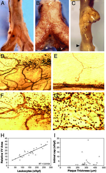
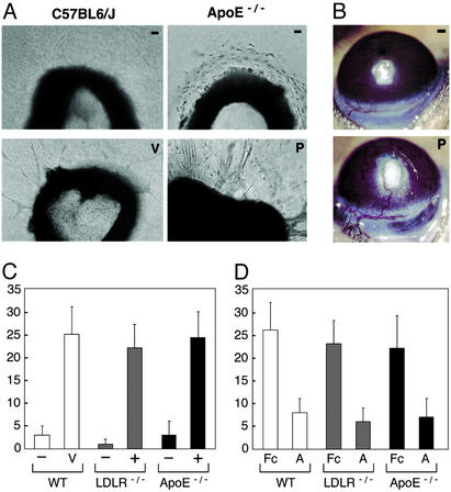
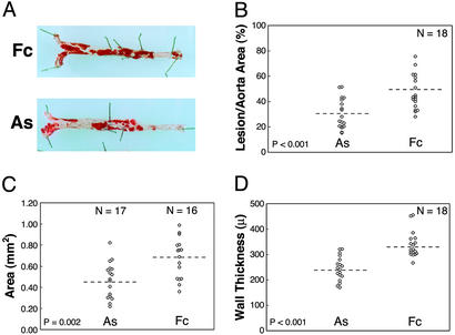
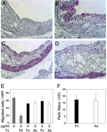
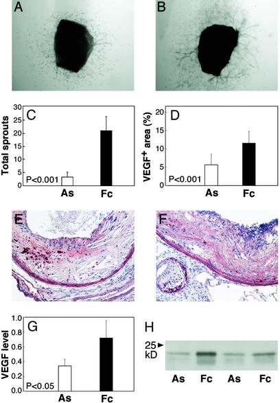
Plaque angiogenesis promotes the growth of atheromas, but the functions of plaque capillaries are not fully determined. Neovascularization may act as a conduit for the entry of leukocytes into sites of chronic inflammation. We observe vasa vasorum density correlates highly with the extent of inflammatory cells, not the size of atheromas in apolipoprotein E-deficient mice. We show atherosclerotic aortas contain activities that promote angiogenesis. The angiogenesis inhibitor angiostatin reduces plaque angiogenesis and inhibits atherosclerosis. Macrophages in the plaque and around vasa vasorum are reduced, but we detect no direct effect of angiostatin on monocytes. After angiogenesis blockade in vivo, the angiogenic potential of atherosclerotic tissue is suppressed. Activated macrophages stimulate angiogenesis that can further recruit inflammatory cells and more angiogenesis. Our findings demonstrate that late-stage inhibition of angiogenesis can interrupt this positive feedback cycle. Inhibition of plaque angiogenesis and the secondary reduction of macrophages may have beneficial effects on plaque stability.


Figures

Figure 1
Figure 1
VV networks associate with regions of intense inflammatory cells in atheromas. (A) Whole-mount CD31 stain of normal aorta from a C57BL6/J mouse shows few VV. The thin aortic wall reveals the CD31+ reaction product (brown) on the aortic endothelium. (B) ApoE−/− mouse aorta is opaque because of an extensive lesion with many adhering VV. (C) One atheroma has VV whereas a nearby advanced lesion (arrow) does not. (D) Clarified flat-mounted aortas demonstrate the CD31+ vascular network associated with some but not all atheromas. (E) VV are absent in regions without lesions. (F) Higher magnification (×400) of lesion in D shows a collection of CD31+ leukocytes surrounding VV. (G) Intense CD31+ mononuclear cells were always associated with VV. (H) VV density (y axis) correlates with the number of CD31+ leukocytes (x axis) in the same region (R2 = 0.8154). (I) Advanced atheromas (n = 23), which all contain intimal capillaries, showed no correlation between maximal intimal depth and the number of capillaries (Spearman rho = 0.28, P = 0.20). (Magnifications: ×5, AC; ×250, D and E; and ×400, F and G.)
Figure 2
Figure 2
Plaque-associated angiogenesis. (A) Aortic explants from C57BL6/J mice produce few sprouts without growth factors (−), but develop maximal sprouts when VEGF (V) or basic fibroblast growth factor is added. Aortic rings from ApoE−/− mice without atheromas (−) produce no sprouts. Aortas that contain an atherosclerotic plaque (P) show marked sprout formation without added stimulators. (B) Plaque-involved aortas (P) stimulate corneal vessels that invade, branch in the plaque, and persist >3 weeks as shown. Aortas without atheromas (−) from ApoE−/− mice do not induce corneal angiogenesis. (Magnifications: ×100, A; ×5, B.) (C) Aortic rings from LDLR−/− and ApoE−/− mice were sorted by the presence (+) or absence (−) of visible lesions. Maximal sprouts developed from lesion-involved aortas, whereas few sprouts formed from aortas without lesions. (D) AS (A, 2 μg/ml) shows 65% inhibition of VEGF-induced sprouts from WT aortas and plaque-induced sprouts from LDLR−/− and ApoE−/− aortas. Total sprouts per ring are shown on the y axis. Bars represent average sprouts per ring ±SD.
Figure 3
Figure 3
AS treatment inhibits lesion progression. (A) Sudan IV-stained aortas isolated from animals representing the median lesion severity for the control (Fc) and AS groups. Atherosclerotic involvement for each mouse was determined by measuring the percent area of Sudan IV+ lesions in the aorta (B), the transverse area of lesions at the aortic sinus (C), and the wall thickness along the inner curve of the aortic arch (D). Statistical comparisons for the AS and Fc groups were based on the independent groups Student's t test. Results from two experiments (n = 18 total mice per group) were statistically significant when analyzed separately and in combination. Dashed lines indicate means.
Figure 4
Figure 4
Reduced plaque macrophages after treatment. Sections from thoracic aorta lesions were stained for macrophages (Mac3) or smooth muscle cells (smooth muscle cell α-actin) shown as the rose color. (A) Fewer macrophages were present in AS lesions compared with Fc (B). Smooth muscle cells were similar in AS (C) and control lesions (D). (Magnifications: ×250.) (E) VEGF-induced migration of human blood monocytes through fibronectin-coated transwell membranes. The mean number of migrated cells per (×400) field was inhibited by Flt-Fc, but not by AS doses that inhibit endothelial cell sprouting. (F) Peritoneal macrophages recruited 24 h (not shown) and 72 h after thioglycollate injection were similar in Fc- and AS-treated mice.
Figure 5
Figure 5
Suppressed angiogenic activity in plaques after in vivo treatment. (A) Sprout formation was determined in four to five rings cut from the thoracic aorta of each treated animal (n = 9 mice per group). Cells migrated out from the AS aortas, but sprouting was markedly reduced and remained suppressed beyond day 12. (B) Atherosclerotic aortas from Fc mice produced similar sprouts as untreated animals (Fig. 2). (C) The mean number of sprouts per animal was significantly reduced in the AS group (P < 0.001). (D) Proximal aortic lesions from AS (E) and control (F) groups show VEGF protein (red) in the media, macrophage-rich areas, and matrix. The percent area of VEGF+ staining in the intima was 2.3-fold decreased in AS compared with control lesions (P < 0.001). (G) Western analysis of VEGF protein in aorta extracts from control and AS-treated mice (n = 7 each group) were 54% reduced in AS animals (P < 0.05). (H) A Western blot represents VEGF levels in plaque extracts. Error bars denote SD. (Magnifications: ×50, A and B; ×100, E and F.)

References

    1. Folkman J. Nat Med. 1995;1:27–31. - PubMed
    1. Ross R. N Engl J Med. 1999;340:115–126. - PubMed
    1. Barger A C, Beeuwkes R D, Lainey L L, Silverman K J. N Engl J Med. 1984;310:175–177. - PubMed
    1. Kaiser M, Younge B, Bjornsson J, Goronzy J J, Weyand C M. Am J Pathol. 1999;155:765–774. - PMC - PubMed
    1. O'Brien K D, McDonald T O, Chait A, Allen M D, Alpers C E. Circulation. 1996;93:672–682. - PubMed

Publication types

MeSH terms

LinkOut - more resources