. 2003 Apr 29;100(9):5240-5.
doi: 10.1073/pnas.0730770100. Epub 2003 Apr 7.

BIR-1, a Caenorhabditis elegans homologue of Survivin, regulates transcription and development

Affiliations

BIR-1, a Caenorhabditis elegans homologue of Survivin, regulates transcription and development

Marta Kostrouchova et al. Proc Natl Acad Sci U S A. .

Abstract

bir-1, a Caenorhabditis elegans inhibitor-of-apoptosis gene homologous to Survivin is organized in an operon with the transcription cofactor C. elegans SKIP (skp-1). Because genes arranged in operons are frequently linked functionally, we have asked whether BIR-1 also functions in transcription. bir-1 inhibition resulted in multiple developmental defects that overlapped with C. elegans SKIP loss-of-function phenotypes: retention of eggs, dumpy, movement defects, and lethality. bir-1 RNA-mediated interference decreased expression of several gfp transgenes and the endogenous genes dpy-7 and hlh-1. Immunoblot analysis revealed decreased phosphoacetylated histones in bir-1 RNA-mediated interference-treated worms. In a heterologous transfection system, BIR-1 augments thyroid hormone-regulated transcription and has an additive effect with SKIP. These results show that BIR-1 functions in the regulation of transcription and development.


Figures
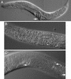
Figure 1
Figure 1
Developmental defects caused by bir-1 inhibition. (A) An affected animal with dumpy (short and thick) phenotype after treatment with bir-1 RNAi. Protrusion of the egg-laying organ is indicated by an arrow. (B) Retention of embryos in the uterus of an adult hermaphrodite treated by bir-1 RNAi. Note the multinucleate embryos. (C) Defective growth of germ line in bir-1 RNAi-treated animals: The germ line makes two turns (arrows), and its growth is elongated. The position of the DTC is marked (arrowhead).
Figure 2
Figure 2
bir-1 RNAi does not influence the expression of CeSKIPgfp. (A) An animal expressing the CeSKIPgfp transgene after treatment with bir-1 RNAi. The expression of CeSKIP is not decreased by bir-1 RNAi. The strong expression is in pharynx (arrowhead), neurons, epidermal cells (arrows) in head region, and in ventral neuronal cord. (B) Distal part of the same animal showing expression of CeSKIPgfp in neurons, epidermal cells, and intestinal muscles. Note the properly developed intestinal muscles expressing CeSKIPgfp (arrow). (C) The presence of Egl phenotype and multinucleate embryos in the same animal as shown in A documenting the developmental defects caused by bir-1 RNAi (Nomarski optics). The arrow indicates the position of the egg-laying organ shown in D. (D) The strong expression of CeSKIPgfp in ventral neuronal cord (arrows) and in egg-laying muscles (arrowhead), which are also normally developed.
Figure 3
Figure 3
The effect of bir-1 inhibition on gene expression. (A) A part of the body of L4 larva showing the expression of elt-2gfp in intestinal cells. The bulbus of pharynx (arrowhead) and normal expression of intestinal cells are marked. (B) Decreased fluorescence of the elt-2gfp transgene is shown in most intestinal cells of a similar larva L4 treated by bir-1 RNAi. The expression of elt-2gfp is not changed in six nuclei in proximal part of the intestine and in one cell in distal part (arrowheads). (C) The normal expression of hlh-1gfp transgene in body wall muscles (arrows) in head and proximal part of the body of a young adult hermaphrodite. (D) Expression of the hlh-1gfp transgene in an animal treated with bir-1 RNAi is decreased markedly.
Figure 4
Figure 4
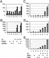
bir-1 augments thyroid hormone-dependent transcription in a heterologous transfection system. (A) Cotransfection of bir-1 enhances basal as well as thyroid hormone-induced transcription in MCF-7 cells. Experiments cotransfected with empty vector are indicated by light columns, and experiments cotransfected with bir-pCDNA3 are indicated by dark columns. Experiments 1–8 are transfected with a reporter (ME-TRE-luc) and control reporter cytomegalovirus-Renilla luciferase: 1–4 are treated with vehicle, 5–8 are treated with thyroid hormone, 1 and 5 are cotransfected with empty vectors pCDNA3, 2 and 6 are cotransfected with human telomerase RNA β1 (hTRβ1), 3 and 7 are cotransfected with mRARα, and 4 and 8 are cotransfected with human telomerase RNA β1 and mRXRα. Results are in relative luciferase units related to control Renilla luciferase expression. BIR-1 (dark columns) augments basal as well as thyroid hormone-induced transcription. (B) BIR-1 has a stimulatory effect on thyroid hormone-induced transcription in MCF-7 cells treated with the histone deacetylase inhibitor TSA. Cells are transfected as described for A. Note that thyroid hormone stimulation of transcription is conserved on TSA background, but the values (related to the same amount of transfected Renilla vector) are ≈20 times higher. (C) The effect of bir-1 and flag-human SKIP are additive in THR-dependent transcription. Experiments 1–8: MCF-7 cells are transfected with a reporter (ME-TRE-luc), control reporter cytomegalovirus-Renilla luciferase, human THRβ1, and mRXRα. Cells are treated with vehicle in the experiments shown in lines 1–4 and with thyroid hormone in 5–8; 1 and 5 are cotransfected with empty vector (pCDNA3), and 2 and 6 are cotransfected with Bir-1 pCDNA3; 3 and 7 are cotransfected with flag-SKIP, and 4 and 8 are cotransfected with Bir-1 pCDNA3 and flag-SKIP. (D and E) The positive effect of BIR-1 and SKIP and their additive effect on thyroid hormone-induced transcription in cos-7 cells. Experiments are as described for C. Cells are treated with vehicle (D) or 10 nM TSA (E). The additive effect of BIR-1 and flag-SKIP is apparent in basal and T3- and T3/TSA-dependent transcription.
Figure 5
Figure 5
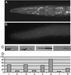
BIR-1 affects phosphoacetylation of histones H3. (A) Immunodetection of S10-P K14-Ac histone H3 in L4 larva. Most nuclei are labeled. (B) Decrease of labeling for S10-P K14-Ac histone H3 in bir-1 RNAi-treated larva L4. (C) Western blot analysis of phosphoacetylated histones H3. Larvae L1–L4 were treated by bir-1 RNAi (lanes 2 and 4) by feeding with bacteria producing dsRNA or carrying empty vector (lanes 1 and 3). Protein extracts were separated by PAGE, blotted on membrane, and immunoanalyzed by using antibody against phosphoacetylated histones H3 [lanes 1 and 2 for S10-P K14-Ac H3 (07-081) and lanes 3 and 4 for K9-Ac S10-P H3 (9711S)]. There is a decrease in phosphoacetylated histone in bir-1 RNAi-treated worms. Lanes 5–8, Western blot analysis of protein extracts from transfected MCF-7 cells. Cells were transfected with bir-1 pCDNA3 (lanes 6 and 8) or empty vector (lanes 5 and 7), harvested 24 h later, and processed for Western blot by using antibodies against S10-P K14-Ac H3 (lanes 5 and 6) and K9-Ac S10-P H3 (lanes 7 and 8). BIR-1 leads to an increase in phosphoacetylated histones H3 (lanes 2 and 4). (D) Densitometric analysis of results.

Similar articles

Cited by

References

    1. Ito M, Roeder R G. Trends Endocrinol Metab. 2001;12:127–134. - PubMed
    1. Felsenfeld G, Clark D, Studitsky V. Biophys Chem. 2000;86:231–237. - PubMed
    1. Jenuwein T, Allis C D. Science. 2001;293:1074–1080. - PubMed
    1. Berger S L, Felsenfeld G. Mol Cell. 2001;8:263–268. - PubMed
    1. Crosio C, Fimia G M, Loury R, Kimura M, Okano Y, Zhou H, Sen S, Allis C D, Sassone-Corsi P. Mol Cell Biol. 2002;22:874–885. - PMC - PubMed

Publication types

LinkOut - more resources