. 2003 Apr 29;100(9):5175-80.
doi: 10.1073/pnas.0534231100. Epub 2003 Apr 7.

Clathrin-coated pits with long, dynamin-wrapped necks upon expression of a clathrin antisense RNA

Affiliations

Clathrin-coated pits with long, dynamin-wrapped necks upon expression of a clathrin antisense RNA

T-G Iversen et al. Proc Natl Acad Sci U S A. .

Abstract

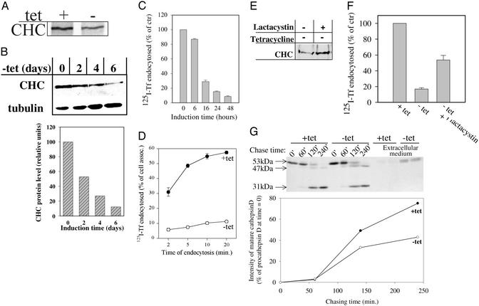
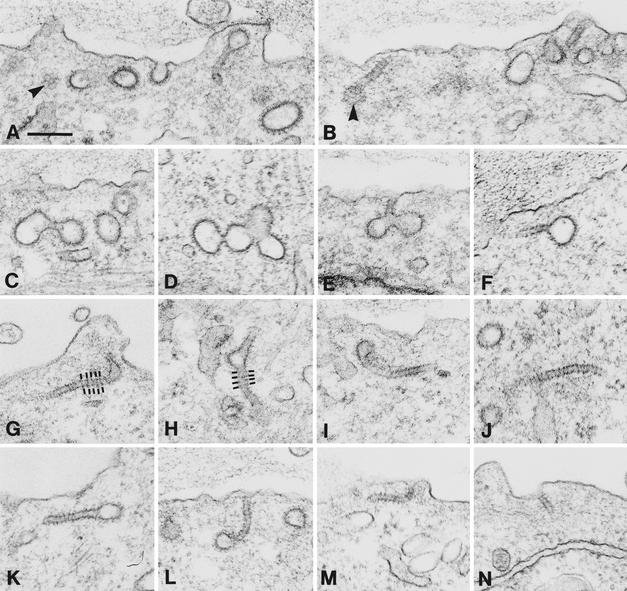
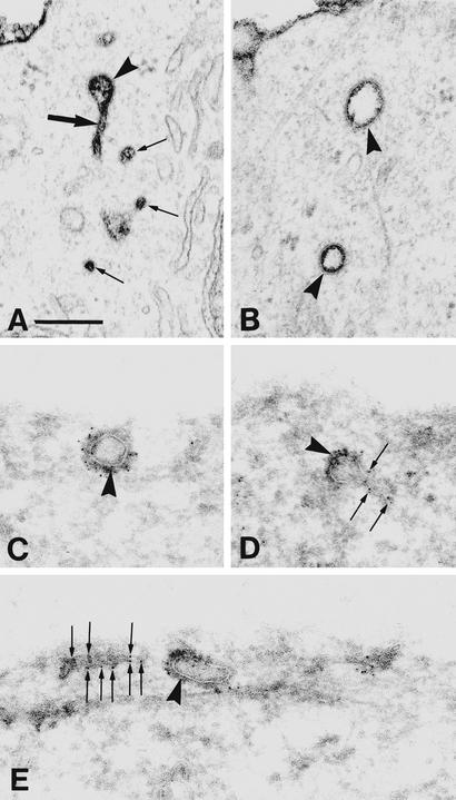
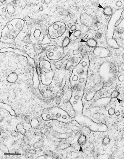
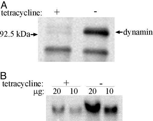
To investigate the role of clathrin in coated vesicle formation, a cell line with inducible expression of clathrin heavy chain (CHC) antisense RNA was produced. After 18 h of CHC antisense RNA expression, the internalization of transferrin was inhibited by 90%. Although the amount of CHC was reduced by only 10%, the frequency of clathrin-coated pits at the cell surface increased by a factor of 3-5, and clathrin-coated structures also accumulated on a pleiomorphic, multivesicular, endosomal compartment. Remarkably, the coated pits were connected to the cell surface by long, tubular necks wrapped by dynamin rings, and the level of dynamin in the CHC antisense RNA-expressing cells was up-regulated 10-fold. In contrast, the amount of several other proteins associated with clathrin coat formation was unaffected. Thus, this study demonstrates that CHC antisense RNA causes accumulation of clathrin-coated pits with dynamin rings around the neck in intact cells not transfected with dynamin mutants, suggesting the existence of a previously uncharacterized functional interplay between clathrin and dynamin.


Figures

Figure 1
Figure 1
Effect of CHC antisense RNA on clathrin and clathrin-dependent trafficking in the BHK21-tTA/anti-CHC cells. (A) Cells grown with or without tet for 2 days were pulse-labeled with [35S]Met/[35S]Cys for 30 min and solubilized. The lysates were immunoprecipitated with goat anti-CHC, and the precipitates were analyzed by SDS/PAGE (7.5% gel) and autoradiography. (B) Cells were grown in the absence of tet for indicated time points and lysed, and equal volumes of the lysates were separated on a 7.5% SDS/PAGE gel. The gel was subjected to Western blot analysis probing with goat anti-CHC antibody and with anti-tubulinα antibody (as internal standard). The film was scanned by densitometry and normalized against tubulin. (C) Cells were grown in the absence of tet for indicated time points and transferred to Hepes medium. 125I-transferrin was added to the cells and endocytosed for 5 min. (D) Cells were grown with or without tet for 2 days. Then the endocytosis of 125I-transferrin for the indicated times was measured. (E) The cells were [35S]Cys/[35S]Met-labeled for 24 h in the presence of tet and further incubated in medium without tet and ± lactacystin for 24 h. CHC was immunoprecipitated with anti-CHC, and the precipitates were analyzed on a 7.5% SDS/PAGE gel. (F) Cells were induced to express CHC antisense RNA for 24 h ± lactacystin, and 125I-transferrin endocytosis during a 5-min incubation at 37°C was measured. (G) Cells were grown with or without tet for 2 days and metabolically labeled. At the indicated chasing times the cells were lysed, and cathepsin D was immunoprecipitated from the lysate and the medium. The precipitates were analyzed on a 12.5% SDS/PAGE gel. The gel then was subjected to autoradiography and densitometric quantification.
Figure 2
Figure 2
Representative examples of clathrin-coated vesicular structures at the cell surface of BHK21-tTA/anti-CHC cells expressing CHC antisense RNA for 2–4 days. The clathrin-coated structures often form clusters. Moreover, they are frequently connected to the cell surface by long tubules. These tubules show a cross-striation (indicated in G and H). Arrowheads in A and B show the characteristic honeycomb-like pattern of tangentially sectioned clathrin-coated pits/vesicles. (Bar = 200 nm.)
Figure 3
Figure 3
(A and B) Representative examples of the peripheral cytoplasm of BHK21-tTA/anti-CHC cells expressing CHC antisense RNA (without tet for 2 days) and fixed in the presence of ruthenium red. Note the ruthenium red-stained clathrin-coated profiles (arrowheads). One such profile is associated with a tubule (large arrow in A); several very small vesicular profiles, which may actually represent cross-sectioned tubules, are also shown (small arrows in A). (C–E) Ultracryosections of the cells expressing CHC antisense RNA (for 3 days). The sections are ImmunoGold-labeled (with Hudy 1 followed by 5-nm gold particles) to identify dynamin, which is present not only on clathrin-coated vesicular profiles (arrowheads) but also on tubular structures (arrows). (Bar = 200 nm.)
Figure 4
Figure 4
BHK21-tTA/anti-CHC cells expressing CHC antisense RNA (without tet for 4 days). Note the numerous clathrin-coated buds (arrowheads) on the pleiomorphic, multivesicular compartment. (Bar = 200 nm.)
Figure 5
Figure 5
Expression of dynamin is increased in the CHC antisense RNA cells. (A) Increased rate of dynamin synthesis. The cells were grown with or without tet for 2 days. Then they were pulse-labeled with [35S]Met/[35S]Cys (30 min) and solubilized. Dynamin was immunoprecipitated and resolved on a 10% SDS/PAGE gel. (B) Northern blot analysis of dynamin 2 mRNA. Total cellular RNA was isolated from cells grown with or without tet for 2 days, and 10 and 20 μg of each RNA sample, as indicated, were loaded onto a 1% agarose/formaldehyde gel and blotted onto a nylon membrane. Hybridization was carried out at 60°C with a labeled cDNA probe for dynamin 2. Band intensities on the blot were quantified with a PhosphorImager.

Comment in

  • Does clathrin pull the fission trigger?
    Di Paolo G, De Camilli P. Di Paolo G, et al. Proc Natl Acad Sci U S A. 2003 Apr 29;100(9):4981-3. doi: 10.1073/pnas.0930650100. Epub 2003 Apr 18. Proc Natl Acad Sci U S A. 2003. PMID: 12704235 Free PMC article. No abstract available.

Similar articles

Cited by

References

    1. Traub L M, Bannykh S I, Rodel J E, Aridor M, Balch W E, Kornfeld S. J Cell Biol. 1996;135:1801–1814. - PMC - PubMed
    1. Stoorvogel W, Oorschot V, Geuze H J. J Cell Biol. 1996;132:21–33. - PMC - PubMed
    1. Johannes L, Goud B. Traffic. 2000;1:119–123. - PubMed
    1. van Dam E M, Stoorvogel W. Mol Biol Cell. 2002;13:169–182. - PMC - PubMed
    1. Raiborg C, Bache K G, Gillooly D J, Madshus I H, Stang E, Stenmark H. Nat Cell Biol. 2002;4:394–398. - PubMed

Publication types