

Biophysical and biological properties of quadruplex oligodeoxyribonucleotides
- PMID: 12682360
- PMCID: PMC153744
- DOI: 10.1093/nar/gkg316
Biophysical and biological properties of quadruplex oligodeoxyribonucleotides
Abstract
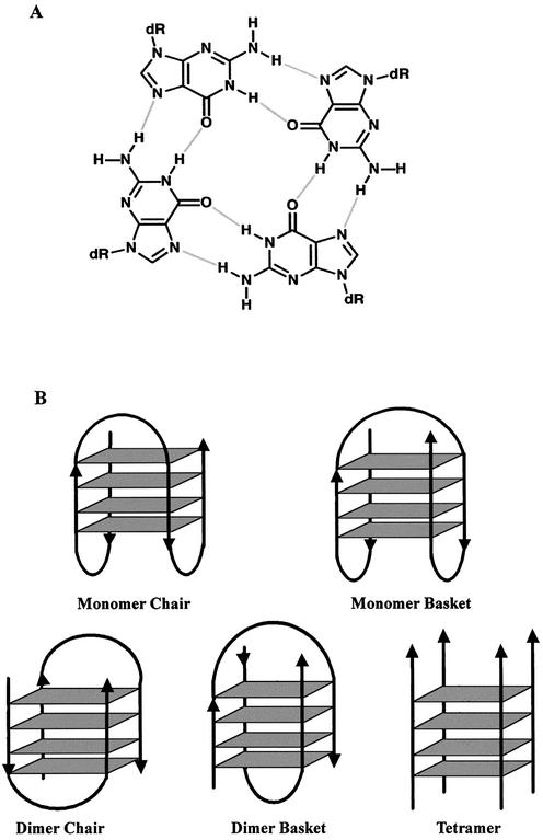
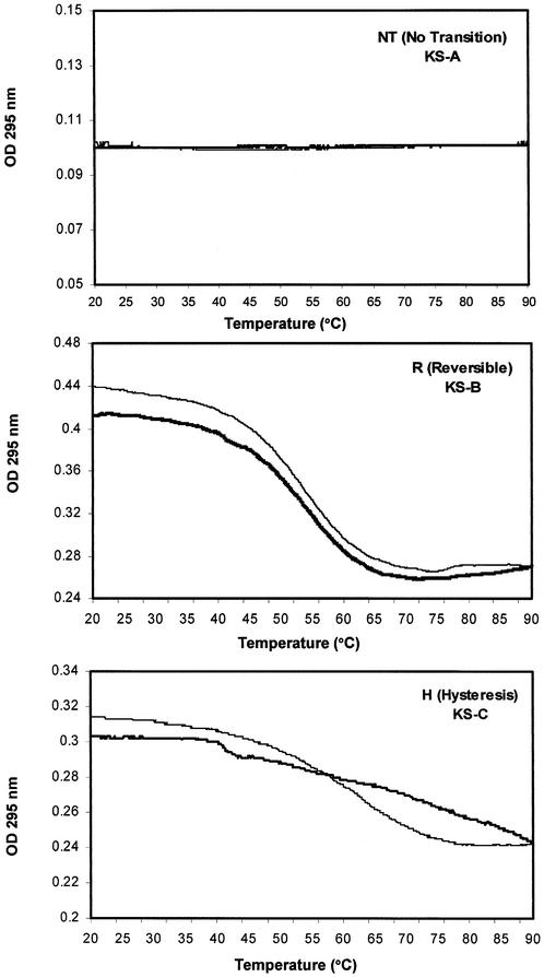
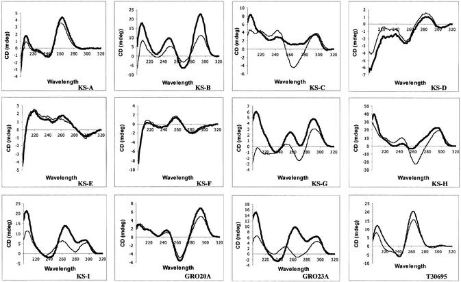
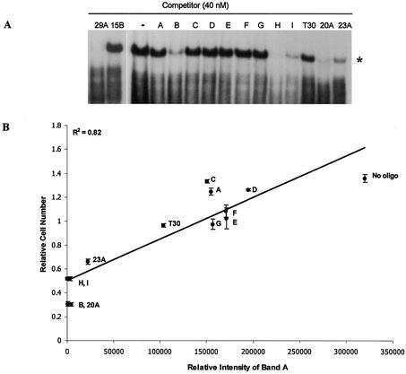
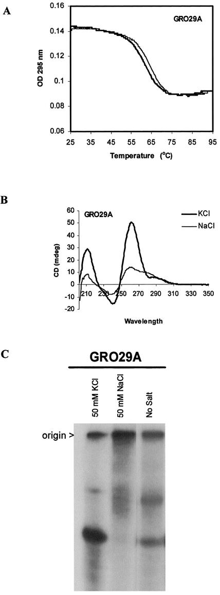
Single-stranded guanosine-rich oligodeoxyribonucleotides (GROs) have a propensity to form quadruplex structures that are stabilized by G-quartets. In addition to intense speculation about the role of G-quartet formation in vivo, there is considerable interest in the therapeutic potential of quadruplex oligonucleotides as aptamers or non-antisense antiproliferative agents. We previously have described several GROs that inhibit proliferation and induce apoptosis in cancer cell lines. The activity of these GROs was related to their ability to bind to a specific cellular protein (GRO-binding protein, which has been tentatively identified as nucleolin). In this report, we describe the physical properties and biological activity of a group of 12 quadruplex oligonucleotides whose structures have been characterized previously. This group includes the thrombin-binding aptamer, an anti-HIV oligonucleotide, and several quadruplexes derived from telomere sequences. Thermal denaturation and circular dichroism (CD) spectropolarimetry were utilized to investigate the stability, reversibility and ion dependence of G-quartet formation. The ability of each oligonucleotide to inhibit the proliferation of cancer cells and to compete for binding to the GRO-binding protein was also examined. Our results confirm that G-quartet formation is essential for biological activity of GROs and show that, in some cases, quadruplex structures formed in the presence of potassium ions are significantly more active than those formed in the presence of sodium ions. However, not all quadruplex structures exhibit antiproliferative effects, and the most accurate factor in predicting biological activity was the ability to bind to the GRO-binding protein. Our data also indicate that the CD spectra of quadruplex oligonucleotides may be more complex than previously thought.
Figures

References
-
- Williamson J.R. (1994) G-quartet structures in telomeric DNA. Annu. Rev. Biophys Biomol. Struct., 23, 703–730. - PubMed
-
- Dempsey L.A., Sun,H., Hanakahi,L.A. and Maizels,N. (1999) G4 DNA binding by LR1 and its subunits, nucleolin and hnRNP D. A role for G–G pairing in immunoglobulin switch recombination. J. Biol. Chem., 274, 1066–1071. - PubMed
-
- Kettani A., Kumar,R.A. and Patel,D.J. (1995) Solution structure of a DNA quadruplex containing the fragile X syndrome triplet repeat. J. Mol. Biol., 254, 638–656. - PubMed
-
- Hanakahi L.A., Sun,H. and Maizels,N. (1999) High affinity interactions of nucleolin with G–G-paired rDNA. J. Biol. Chem., 274, 15908–15912. - PubMed
Publication types
MeSH terms
Substances
LinkOut - more resources
Full Text Sources
Other Literature Sources
