. 2003 Mar;138(6):1107-18.
doi: 10.1038/sj.bjp.0705161.

Erythropoietin fosters both intrinsic and extrinsic neuronal protection through modulation of microglia, Akt1, Bad, and caspase-mediated pathways

Affiliations

Erythropoietin fosters both intrinsic and extrinsic neuronal protection through modulation of microglia, Akt1, Bad, and caspase-mediated pathways

Zhao Zhong Chong et al. Br J Pharmacol. 2003 Mar.

Abstract

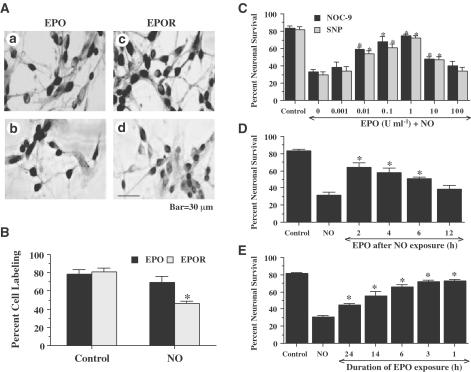
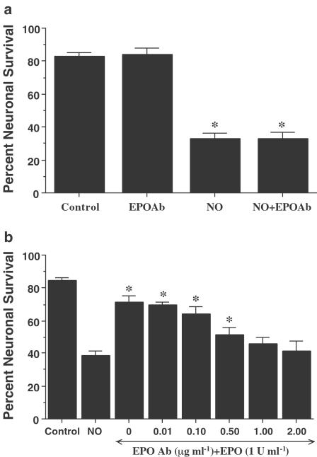
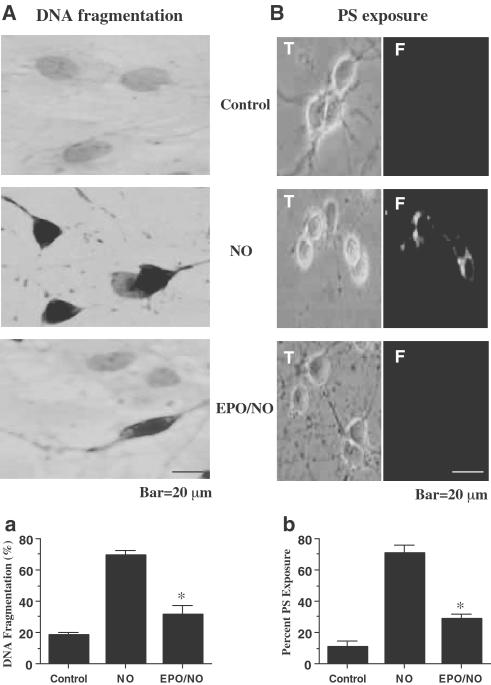
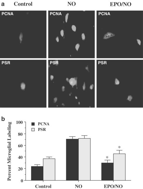
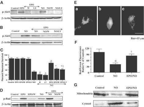
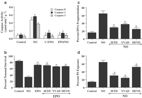
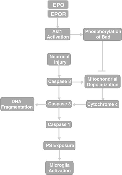
1. Erythropoietin (EPO) plays a significant role in the hematopoietic system, but the function of EPO as a neuroprotectant and anti-inflammatory mediator requires further definition. We therefore examined the cellular mechanisms that mediate protection by EPO during free radical injury in primary neurons and cerebral microglia. 2. Neuronal injury was evaluated by trypan blue, DNA fragmentation, phosphatidylserine (PS) exposure, Akt1 phosphorylation, Bad phosphorylation, mitochondrial membrane potential, and cysteine protease activity. Microglial activation was assessed through proliferating cell nuclear antigen and PS receptor expression. 3. EPO provides intrinsic neuronal protection that is both necessary and sufficient to prevent acute genomic DNA destruction and subsequent membrane PS exposure, since protection by EPO is completely abolished by cotreatment with an anti-EPO neutralizing antibody. 4. Extrinsic protection by EPO is offered through the inhibition of cerebral microglial activation and the suppression of microglial PS receptor expression for the prevention of neuronal phagocytosis. In regards to microglial chemotaxis, EPO modulates neuronal poptotic membrane PS exposure necessary for microglial activation primarily through the regulation of caspase 1. 5. EPO increases Akt1 activity, phosphorylates Bad, and maintains neuronal nuclear DNA integrity through the downstream modulation of mitochrondrial membrane potential, cytochrome c release, and caspase 1, 3, and 8-like activities. 6. Elucidating the intrinsic and extrinsic protective pathways of EPO that mediate both neuronal integrity and inflammatory microglial activation may enhance the development of future therapies directed against acute neuronal injury.


Figures

Figure 1
Figure 1
Protection by EPO is concentration and time dependent during diminished expression of the EPOR during NO exposure. (A) Primary hippocampal neuronal cultures were subjected to immunocytochemical detection for EPO (Aa, Ab) and the EPOR (Ac, Ad) by using a rabbit primary polyclonal anti-EPO and anti-EPOR antibodies. For EPO and EPOR detection, representative images are displayed for control cells (untreated neurons) (Aa, Ac) and for cells 24 h following exposure to the NO donor NOC-9 (300 μM) in the adjacent panels (Ab, Ad). (B) Quantitation of the percentage of neurons expressing EPO or the EPOR at 24 h following exposure to either NOC-9 (300 μM) or SNP (300 μM) is shown (*P<0.01 vs untreated control). (C) Neurons were pretreated with EPO (0.001–100 U ml−1) 1 h prior to exposure to a NO donor (NOC-9 or SNP, 300 μM) and cell survival was assessed 24 h later. Protection of EPO against NO toxicity was evident in cultures with EPO (0.01–10 U ml−1) when compared with cultures exposed to NO alone (*P<0.01 vs NO treated alone). (D) Protection of EPO was evident in post-treatment paradigms during NO toxicity. Neurons were treated with EPO (1 U ml−1) at 2, 4, 6, and 12 h following NO exposure (NOC-9 or SIN-1, 300 μM). Post-treatment with EPO at 2, 4, and 6 h following NO exposure increased neuronal survival significantly 24 h following NO exposure (*P<0.01 vs NO treated alone). (E) EPO (1 U ml−1) was pre-administered at 1, 3, 6, 14, and 24 h prior to NO exposure and neuronal survival was assessed 24 h following NO application (NOC-9 or SNP, 300 μM). Administration of EPO at 1, 3, and 6 h prior to NO exposure generated the highest levels of neuronal survival, but EPO applications provided at 14 and 24 h resulted in decreased efficacy for neuroprotection by EPO (*P<0.01 vs NO treated alone; †P<0.01 vs 24 h pretreated group). In (B, D, E), to simplify the figures, the results for the two NO donors were combined.
Figure 2
Figure 2
EPO is necessary and sufficient for neuronal protection during NO exposure. (a) To examine the ability of the EPO Ab to alter neuronal viability during NO exposure, EPO Ab (2 μg ml−1) was applied 1 h prior to increasing concentrations of a NO donor (NOC-9 or SNP, 300 μM). Neuronal survival was assessed 24 h following NO application. Administration of EPO Ab alone was not toxic. No significant changes in neuronal survival were observed following application of the EPO Ab when compared to cultures treated with NO alone (*P<0.01 vs untreated control). (b) Increasing concentrations of the EPO Ab (0.01–2.00 μg ml−1) were applied to neuronal cultures in conjunction with EPO (1 U ml−1) for 1 h prior to NO exposure (NOC-9 or SNP, 300 μM). Neuronal survival was assessed 24 h following NO application. Protection by EPO against NO toxicity was attenuated or abolished during applications of EPO Ab (0.50, 1.00, and 2.00 μg ml−1) (*P<0.01 vs NO treated alone). In (a) and (b), to simplify the figures, the results for the two NO donors were combined.
Figure 3
Figure 3
EPO prevents DNA fragmentation and externalization of membrane PS residues in neurons. (A) Neurons were exposed to NO (NOC-9 or SNP, 300 μM) and DNA fragmentation was determined 24 h later using the TUNEL assay. Pretreatment with EPO (1 U ml−1) decreased DNA fragmentation significantly during NO exposure (*P<0.01 vs NO, panel Aa). (B) Neurons were labeled with annexin V PE to visualize PS exposure 24 h following exposure to NO (NOC-9 or SNP, 300 μM) and were imaged using transmitted (T) light and corresponding fluorescence (F) images of the same microscopy field. Pretreatment with EPO (1 U ml−1) 1 h prior to NO significantly prevented membrane PS externalization (*P<0.01.3 vs NO, panel Bb). In (Aa) and (Bb), to simplify the figures, the results of the two NO donors were combined. In all cases, control = untreated neurons.
Figure 4
Figure 4
EPO protects against microglial activation and microglial PSR expression during NO exposure. Pure microglial cultures were treated for 3 h with media with or without 1 h EPO (1 U ml−1) pretreatment that had been exposed to NO (NOC-9 or SNP, 300 μM) 24 h prior. (a) A representative image illustrates that PCNA or PSR expression was significantly increased in microglia treated with media from NO exposed neurons. In contrast, PCNA expression or PSR expression was significantly less in microglia treated with media from EPO (1 U ml−1) and NO-treated neurons. (b) PCNA expression or PSR expression in microglia treated with media from NO-exposed neurons was significantly increased, but PCNA or PSR expression was significantly diminished in microglia treated with media from EPO (1 U ml−1) and NO-treated neurons EPO/NO vs NO, *P<0.01). To simplify the figures, the results of the two NO donors were combined. Control = cultures without NO exposure.
Figure 5
Figure 5
Neuronal protection by EPO is mediated by the activation of Akt1, phosphorylation of Bad, and the prevention of mitochondrial membrane depolarization and cytochrome c release. In (A) and (B), equal amounts of neuronal protein extracts (50 μg per lane) were immunoblotted with antiphospho-Akt1 (p-Akt1, active Akt1, Ser 473) antibody. Exposure to EPO (1 U ml−1) or NO significantly increased p-Akt1 expression. Application of the PI-3K inhibitor wortmannin (100 nM) or LY294002 (LY) (10 μM) was sufficient to block the expression of active p-Akt1 in the presence of EPO during NO (NOC-9, 300 μM) exposure. (C) At a concentration that blocks activation of p-Akt1 during NO administration (NOC-9 or SNP, 300 μM), wortmannin (100 nM) or LY294002 (10 μM) applied 1 h prior to NO significantly reduced the protective capacity of EPO (1 U ml−1) during NO exposure (*P<0.01 vs NO; †P<0.01 vs EPO). (D) Equal amounts of neuronal protein extracts (50 μg per lane) were immunoblotted with antiphosphorylated Bad (p-Bad, Ser 136) antibody. Exposure to EPO (1 U ml−1) or NO significantly increased p-Bad expression. EPO application further increased phosphorylation of Bad during NO exposure. Application o of the PI-3K inhibitor wortmannin (100 nM) was sufficient to block the expression of p-Bad in the presence of EPO during NO (NOC-9, 300 μM) exposure. (E, F) Exposure to NO (NOC-9, 300 μM) produced a significant decrease in the red/green fluorescence intensity ratio using a cationic membrane potential indicator JC-1 within 3 h when compared with untreated control cultures, suggesting that NO results in mitochondrial membrane depolarization. Application of EPO (1 U ml−1) 1 h prior to NO exposure significantly increased the red/green fluorescence intensity of neurons, indicating that mitochondrial permeability transition pore membrane potential was restored to baseline (E,F). (G) A representative Western blot with equal amounts of mitochondrial or cytosol protein extracts (50 μg per lane) were immunoblotted demonstrating that application of EPO (1 U ml−1) significantly prevented cytochrome c release from mitochondria during NO exposure. In (C) and (F), to simplify the figures, the results of the two NO donors were combined. In all cases, control=untreated neurons.
Figure 6
Figure 6
EPO protects neurons from injury through the modulation of caspase 8, caspase 1, and caspase 3-like activities. (a) Neurons were exposed to NO (NOC-9 or SNP, 300 μM) and caspase 8-, caspase 1-, and caspase 3-like activities were assessed 12 h later through their respective colorimetric substrates. Pretreatment with EPO (1 U ml−1) or the caspase inhibitors (C-I, 50 μM) for caspase 8 (IETD), caspase 1 (YVAD), and caspase 3 (DEVD) 1 h prior to NO significantly inhibited the increase in the activity of caspase 8, caspase 1, and caspase 3 induced by NO (*P<0.01 vs NO). (b) Neurons were pretreated with EPO (1 U ml−1) alone or in combination with an inhibitor of caspase 8 (IETD, 50 μM), caspase 1 (YVAD, 50 μM), or caspase 3 (DEVD, 50 μM). No enhanced or synergistic protection was observed during the application of each caspase inhibitor combined with EPO when compared with cultures exposed to EPO and NO alone. (c) Neurons were pretreated with a caspase 8 inhibitor (IETD, 50 μM), a caspase 1 inhibitor (YVAD, 50 μM), or a caspase 3 inhibitor (DEVD, 50 μM) 1 h prior to NO (NOC-9 or SNP, 300 μM) and DNA fragmentation with TUNEL was determined 24 h following NO exposure (*P<0.01 vs NO). (d) Neurons were pretreated with a caspase 8 inhibitor (IETD, 50 μM), a caspase 1 inhibitor (YVAD, 50 μM), or a caspase 3 inhibitor (DEVD, 50 μM) 1 h prior to NO (NOC-9 or SNP, 300 μM) and membrane PS exposure with annexin V PE was determined 24 h following NO exposure (*P<0.01 vs NO). In all cases, to simplify the figures, the results of the two NO donors were combined and control = untreated neurons.
Figure 7
Figure 7
EPO prevents neuronal injury through a series of pathways that involve Akt1, Bad, and cysteine protease activity. Following neuronal injury, both caspase 8 and Bad can lead to the depolarization of the mitochondrial membrane resulting in the release of cytochrome c. Concurrently, caspase 8 also can activate caspase 3 to precipitate DNA fragmentation and potentially activate caspase 1 to yield membrane PS externalization, microglial activation, and the phagocytic destruction of neurons. The prevention of neuronal apoptosis and microglial phagocytosis by EPO following its binding to the EPOR during NO.6 exposure can occur through cellular pathways that involve enhanced Akt1 activity, Bad phosphorylation, and the maintenance of mitochondrial membrane stability. Alternatively, EPO may act directly upon cytochrome c, caspase 8, caspase 3, or caspase 1 to promote neuronal survival during toxic insults.

References

    1. ACS G., ACS P., BECKWITH S.M., PITTS R.L., CLEMENTS E., WONG K., VERMA A. Erythropoietin and erythropoietin receptor expression in human cancer. Cancer Res. 2001;61:3561–3565. - PubMed
    1. ANDERSON I., ADINOLFI C., DOCTROW S., HUFFMAN K., JOY K.A., MALFROY B., SODEN P., RUPNIAK H.T., BARNES J.C. Oxidative signalling and inflammatory pathways in Alzheimer's disease. Biochem. Soc. Symp. 2001;67:141–149. - PubMed
    1. BAL-PRICE A., BROWN G.C. Nitric-oxide-induced necrosis and apoptosis in PC12 cells mediated by mitochondria. J. Neurochem. 2000;75:1455–1464. - PubMed
    1. BERNAUDIN M., MARTI H.H., ROUSSEL S., DIVOUX D., NOUVELOT A., MACKENZIE E.T., PETIT E. A potential role for erythropoietin in focal permanent cerebral ischemia in mice. J. Cerer. Blood Flow Metab. 1999;19:643–651. - PubMed
    1. BLUME-JENSEN P., JANKNECHT R., HUNTER T. The kit receptor promotes cell survival via activation of PI 3-kinase and subsequent Akt-mediated phosphorylation of Bad on Ser136. Curr. Biol. 1998;8:779–782. - PubMed

Publication types

MeSH terms