. 2003 Apr;2(2):295-305.
doi: 10.1128/EC.2.2.295-305.2003.

Transcriptional silencing of an amoebapore gene in Entamoeba histolytica: molecular analysis and effect on pathogenicity

Affiliations

Transcriptional silencing of an amoebapore gene in Entamoeba histolytica: molecular analysis and effect on pathogenicity

Rivka Bracha et al. Eukaryot Cell. 2003 Apr.

Abstract

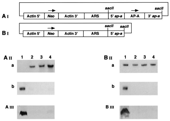
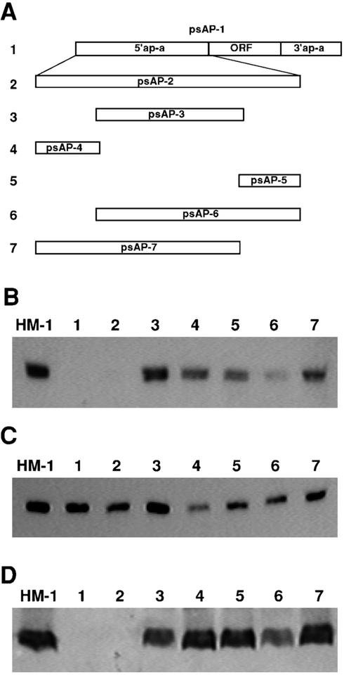
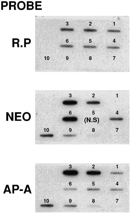
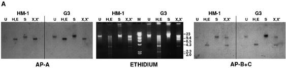
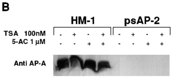
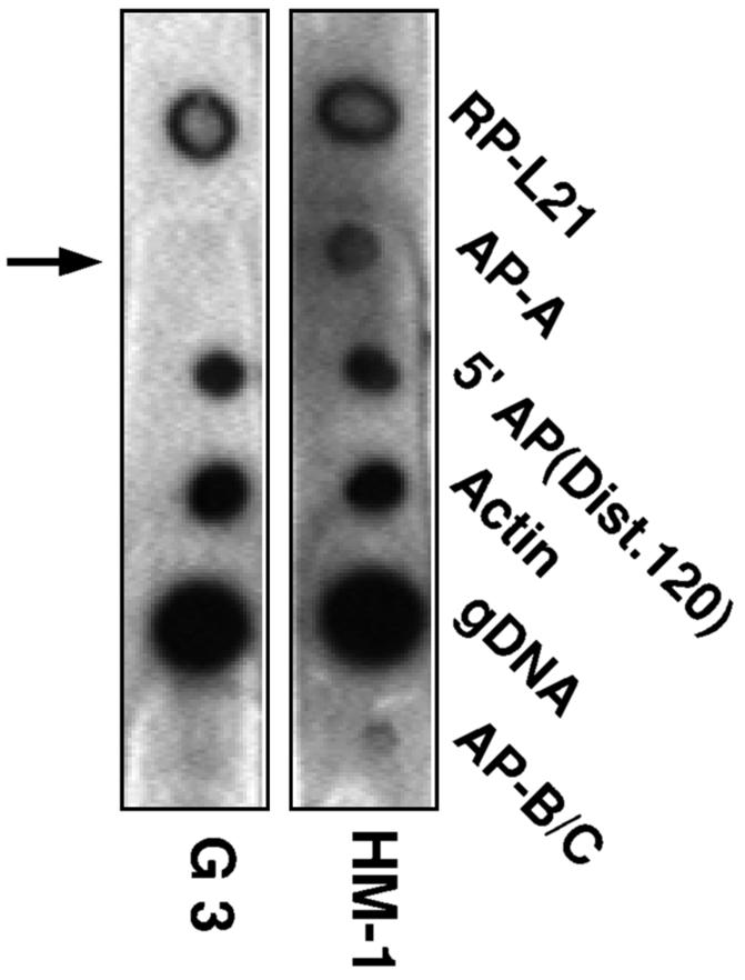
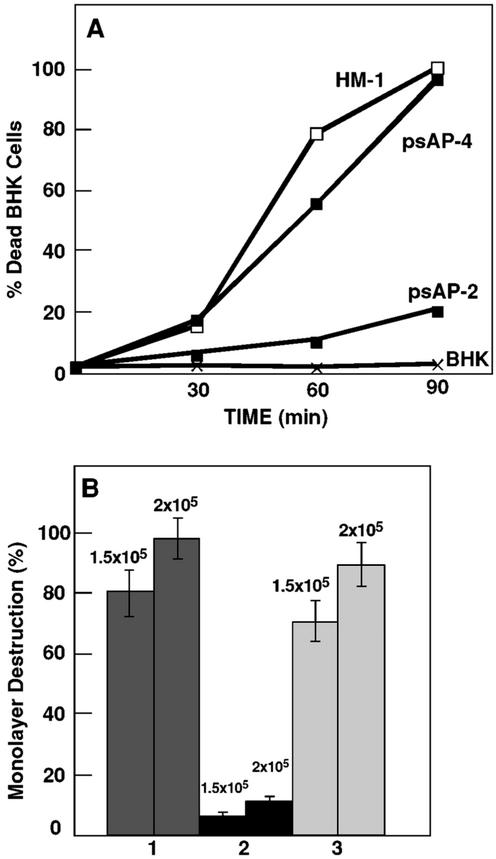
Transcriptional silencing of the gene coding for amoebapore A (AP-A) was observed when trophozoites of Entamoeba histolytica were transfected with a hybrid plasmid construct containing the ap-a gene flanked by the upstream and downstream segments of the original Ehap-a gene. Transfectants were totally devoid of ap-a transcript and AP-A protein. An identical silencing effect was observed upon transfection with a plasmid that contained only the 5' upstream region of ap-a. Removal of the selecting antibiotic enabled the isolation of plasmidless clones, which retained in their progeny the silenced phenotype. E. histolytica cells were able to overexpress ap-a when transfected with a plasmid containing the gene flanked by the 5' and 3' regions of the EhRP-L21 gene. This plasmid, however, could not express ap-a in the retransfected, cloned trophozoites lacking AP-A. This is the first report of gene silencing in E. histolytica, and the mechanism appears to belong to transcriptional gene silencing and not to posttranscriptional gene silencing. This conclusion is based on the following results: (i) silencing was achieved by transfection of homologous 5' flanking sequences (470 bp of the Ehap-a gene), (ii) transcription initiation of Ehap-a was found to be blocked, and (iii) short double-stranded RNA fragments of the ap-a coding and noncoding sequences were not detected. Trophozoites lacking AP-A are nonpathogenic and impaired in their bacteriolytic capability.


Figures

FIG. 1.
FIG. 1.
(AI) psAP-1 plasmid in which the ap-a gene, including sequences from its upstream and downstream regulatory regions, was inserted into a SacII site of the pEhActNeo shuttle vector (1, 36). ARS, autonomous replication sequence. (AII) Northern blot analysis of amoebic RNA extracted from the following sources: parent strain HM-1:IMSS (lane 1) and psAP-1 transfectants grown in the presence of 6 (lane 2), 12 (lane 3), and 24 (lane 4) μg of G418 ml−1. The DNA probes used were Neo (AIIa) and ap-a (AIIb). (AIII) Western blot of SDS-PAGE reacted with anti AP-A antibodies. Lanes depict results for the same cultures as described above. (BI) psAP-2 plasmid in which only the 470 bp of the 5′ flanking region was cloned in the same vector as described above. (BII) Northern analysis of psAP-2 transfectant. Lane 1, HM-1:IMSS parent strain; lanes 2, 3, and 4, psAP-2 transfectants grown in the presence of 6 (lane 2), 12 (lane 3), and 24 (lane 4) μg of G418 ml−1. The DNA probes used were ribosomal protein (BIIa) and ap-a (BIIb). (BIII) Western blot of SDS-PAGE reaction with anti AP-A. Lanes depict results for the same cultures as described above.
FIG. 2.
FIG. 2.
(A) Schematic map of the plasmids used to examine the regions of the 5′ upstream 470-bp segment, which may be responsible for silencing. Using the primers described in Table 1, plasmids psAP-2 to psAP7 were derived from plasmid psAP-1. (B) Northern blots probed with ap-a gene. (C) Northern blot probed with the ribosomal protein gene EhRP-L21. (D) Western blot reacted with anti-AP-A antibodies and horseradish peroxidase anti-rabbit antibodies. Lane HM-1, control parent strain; lane 1, psAP-1; lane 2, psAP-2; lane 3, psAP-3; lane 4, psAP-4; lane 5, psAP-5; lane 6, psAP-6; lane 7, psAP-7.
FIG. 3.
FIG. 3.
Quantitation of plasmid copies in DNA of 5 × 10⁵ transfected amoeba from the following sources: HM-1 (slot 1); psAP-1 grown with 6 (slot 2) and 100 (slot 3) μg of G418 ml−1; psAP-2 grown with 6 μg of G418 ml−1 (slot 4); and psAP-4 grown with 6 (slot 5) and 100 (slot 6) μg of G418 ml−1. Slots 7, 8, 9, and 10 show samples from plasmid psAP-1 at a multiplicity (compared to the genomic DNA) of 0.8 (slot 7), 2 (slot 8), 8 (slot 9), and 20 (slot 10). Each blot was probed with different labeled probes. (R.P) With ribosomal protein L-21, all samples showed similar levels and the plasmid control showed no signal. (NEO) The nontransfected HM-1 strain (slot 1) showed no signal. Differences were seen between the samples grown at 6 μg of G418 ml−1 (slots 2 and 4) and those grown at 100 μg ml−1 (slots 3 and 6), and psAP-1 plasmid signal levels were relative to the amount spotted. (AP-A) Only samples 2 (slot 2) and 3 (slot 3), containing psAP-1 plasmid, had extra copies of the ap-a gene compared to those of HM-1. The other transfectant showed the same number of copies as the control strain (slot 1). N.S, no sample in the slot.
FIG. 4.
FIG. 4.
Characterization of the plasmidless AP-A lacking trophozoite clone G3. (A) Northern blots of various transfectants as well as plasmidless trophozoites probed with a variety of probes. Lanes 1, HM-1:IMSS nontransfected parent strain; lane 2, transfectant psAP-2; lane 3, plasmidless clone G3 isolated from cultures of transfectant psAP-2 grown in the absence of G418; lane 4, clone G3 retransfected with plasmid pA7; lane 5, clone G3 retransfected with plasmid pTS1, which contains the CAT gene (35); lane 6, strain HM-1:IMSS transfected with plasmid pTS1. Probes were as marked. RP-L21 served as loading control. The AP-A probe revealed a complete absence of transcript in lanes 2, 3, and 4. Probe AP-B+C contained in tandem the sequences of genes ap-b and ap-c and was used for their detection. The result revealed a lower level of transcription. Probe 5′AP(Dist. 120), which consists of the distal 120 bp of the 470-bp 5′ flanking element of gene Ehap-a, hybridized to an unidentified transcript. (B) Western blot of SDS-PAGE results for lysates of trophozoites reacted with anti AP-A. Lanes contain the same samples as described above. Lanes 2, 3, and 4 showed no detectable AP-A. (C1) pA7 plasmid in which the ap-a gene is flanked by the 5′ (5′gLE3) and 3′ (3′gLE3) elements of the EhRP-L21 gene (13). ARS is the sequence containing the autonomous replication element. (C2 and C3) Northern and Western blots, respectively, of strain HM-1:IMSS and of HM-1:IMSS trophozoites transfected with plasmid pA7 and grown with G418 at the indicated concentrations.
FIG. 5.
FIG. 5.
(A) Southern blots of restriction digests of DNA samples from nontransfected parent strain HM-1:IMSS or plasmidless, AP-A-lacking clone G3. Samples were undigested (U) or digested with HindIII and EcoRI (H,E), SalI (S), or XhoI and XbaI (X,X′). (Middle panel) Ethidium-stained agarose gel. (Left and right panels) Blots were probed under stringent conditions with probes specific for ap-a and with a probe containing in tandem the sequences of ap-b and ap-c genes, respectively. (B) Western blot analysis of AP-A with trophozoites grown for 24 h with trichostatin A (TSA) (100 nM), 5′aza-2′deoxycytosine (5-AC) (1 μM), or with both compounds. The amount of AP-A in the lysates was monitored with antibodies against AP-A (Anti AP-A). The trophozoites were from strain HM-1:IMSS (HM-1) and psAP2 (as marked).
FIG. 5.
FIG. 5.
(A) Southern blots of restriction digests of DNA samples from nontransfected parent strain HM-1:IMSS or plasmidless, AP-A-lacking clone G3. Samples were undigested (U) or digested with HindIII and EcoRI (H,E), SalI (S), or XhoI and XbaI (X,X′). (Middle panel) Ethidium-stained agarose gel. (Left and right panels) Blots were probed under stringent conditions with probes specific for ap-a and with a probe containing in tandem the sequences of ap-b and ap-c genes, respectively. (B) Western blot analysis of AP-A with trophozoites grown for 24 h with trichostatin A (TSA) (100 nM), 5′aza-2′deoxycytosine (5-AC) (1 μM), or with both compounds. The amount of AP-A in the lysates was monitored with antibodies against AP-A (Anti AP-A). The trophozoites were from strain HM-1:IMSS (HM-1) and psAP2 (as marked).
FIG. 6.
FIG. 6.
Hybridization of blots with radiolabeled nascent RNA extracted from nuclear run-on experiments of control nontransfected HM-1:IMSS trophozoites and AP-A-lacking trophozoites of clone G3. E. histolytica DNA samples on the blots were prepared by PCR on E. histolytica DNA and included the ribosomal protein L21 (RP-L21), the ap-a gene (AP-A), a 5′ flanking fragment (120 bp) of the ap-a gene (bp −470 to −350) [5′ AP(Dist.120)], actin, genomic DNA (gDNA), and DNA of tandemly linked ap-b and ap-c genes (AP-B/C). 300 ng of each DNA was spotted and denatured prior to hybridization.
FIG. 7.
FIG. 7.
(A) Cytotoxic activity. The mortality rates of BHK cells incubated in suspension with freshly harvested trophozoites of strain HM-1:IMSS, psAP-2 transfectants, and psAP-4 transfectants (which served as control) are shown. Both transfectants were grown with 6 μg of G418 ml−1. The number of trypan blue-stained BHK cells was monitored as a function of incubation time. The percentage of BHK cells that incorporated the dye in the absence of trophozoites remained low (<4%) during the period tested. (B) Cytopathic activity of different trophozoite cultures of HM-1:IMSS (lane 1), psAP-2 transfectants (lane 2), and psAP-4 control transfectants (lane 3). Both transfectants were grown with 6 μg of G418 ml−1. Two concentrations (1.5 × 10⁵ and 2 × 10⁵) of trophozoites were added per well for each type of trophozoite; experiments were repeated in triplicate. Bars represent standard errors. Statistical significance was determined by single-tailed t testing (P < 0.05).
FIG. 8.
FIG. 8.
The total amount of 14C-labeled E. coli cells which attached and became ingested by the trophozoites, as determined after removal of the nonattached bacteria by Percoll gradients (10), is depicted using filled bars. The percentage of radiolabeled bacteria (out of the total attached) which solubilized after treatment of trophozoite samples with Triton x-100 is depicted using empty bars. Left bar pair, strain HM-1:IMSS; center bar pair, transfectant psAP-2, right bar pair, transfectant psAP-4.
FIG. 9.
FIG. 9.
Confocal fluorescent microscopy of trophozoites that ingested GFP-labeled E. coli cells. Trophozoites were associated with the bacteria for 1 h, after which they were harvested and washed. A portion of the total amount of trophozoites was then fixed in formaldehyde, and the other portion was resuspended in fresh medium for further incubation (23 h) in the presence of antibiotics (Claforan) to prevent the growth of the noningested bacteria. After that period, the trophozoites were harvested, washed, and fixed in formaldehyde. The panel labeled “Super imposed” combines the image obtained by phase contrast (Nomarski optics) with the fluorescent image to show the bacterial location in the vacuoles.

References

    1. Alon, R. N., R. Bracha, and D. Mirelman. 1997. Inhibition of expression of the lysine-rich 30 kDa surface antigen of Entamoeba dispar by the transcription of its antisense RNA. Mol. Biochem. Parasitol. 90:193-201. - PubMed
    1. Ankri, S., F. Padilla-Vaca, T. Stolarsky, L. Koole, U. Katz, and D. Mirelman. 1999. Antisense inhibition of expression of the light subunit (35 kDa) of the Gal/GalNAc lectin complex inhibits Entamoeba histolytica virulence. Mol. Microbiol. 33:327-337. - PubMed
    1. Ankri, S., T. Stolarsky, R. Bracha, F. Padilla-Vaca, and D. Mirelman. 1999. Antisense inhibition of expression of cysteine proteinases affects Entamoeba histolytica-induced formation of liver abscess in hamsters. Infect. Immun. 67:421-422. - PMC - PubMed
    1. Ankri, S., T. Stolarsky, and D. Mirelman. 1998. Antisense inhibition of expression of cysteine proteinases in Entamoeba histolytica does not affect cytopathic or hemolytic activity but inhibits phagocytosis. Mol. Microbiol. 28:777-785. - PubMed
    1. Arhets, P., J. C. Olivo, P. Gounon, P. Sansonetti, and N. Guillen. 1998. Virulence and functions of myosin II are inhibited by overexpression of light meromyosin in Entamoeba histolytica. Mol. Biol. Cell 9:1537-1547. - PMC - PubMed

Publication types

MeSH terms